Application
XploRA INV
Overview
The XploRA INV combines the exclusive automation features and small footprint of the standard XploRA™ Raman microscope with the unique sampling capabilities of an inverted microscope, especially important for demanding biological applications. These applications include:
- cell research
- disease detection
- characterisation of drug-cell interactions
- pharmaceutical verification of intercellular activities
In addition, the inverted geometry allows easy incorporation of AFM units for combined Raman-AFM and TERS (Tip Enhanced Raman Spectroscopy) for highest spatial resolution sample visualisation.
The open structure of the XploRA INV also permits the use of virtually all options and add-ons for inverted microscopes, including micro manipulators and “optical tweezers”, specific enclosures for cell applications, etc.

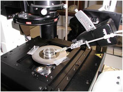
XploRA INV sample stage equipped with micro-injector
The XploRA INV also offers the option to integrate the unique scanning capabilities of HORIBA’s patented DuoScan for rapid spectroscopic multichannel Raman imaging, as well as the new 3D FCI (Fast Confocal Imaging) module, which allows ultra-fast fluorescence imaging for visualisation and verification.


White light image (left), fluorescence FCI image (center) and hyperspectral fluorescence image (right) images of a bovine embryo after staining with Nile Red, acquired on the XploRA INV. The hyperspectral image illustrates the stain in different polarity states. Data courtesy of Prof. Igor Chourpa, Université de Tours, France.
Manufactured by HORIBA Scientific


