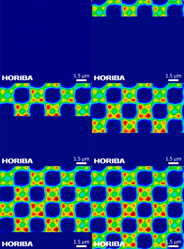
Imagerie Raman ultrarapide SWIFT de deuxième génération, montrant l'acquisition en temps réel d'une image Raman hyperspectrale sur un dispositif semi-conducteur structuré, avec 40 200 spectres acquis en moins de 50 secondes.
Avantages de l'imagerie Raman rapide :
- Diminution du temps d'acquisition de plusieurs ordres de grandeur sans compromis sur la qualité d'image.
- Possibilité d'analyser des macro-zones (centimètres) avec la micro-résolution.
- Possibilité d'obtenir des images Raman haute définition de plusieurs mégapixels dans une seule cartographie Raman rapide, permettant de combiner une vue d'ensemble et une vue haute résolution détaillée au sein de la même expérience.
- Possibilité d'acquérir des images de volumes confocaux 3D contenant de nombreuses données dans des délais réalistes pour interroger la structure interne d'un échantillon.
- Imagerie Raman résolue dans le temps : l'acquisition d'une image complète en quelques secondes seulement permet désormais de visualiser des réactions chimiques, telles que le durcissement des polymères, qui se produisent en quelques minutes/heures.
La technologie SWIFT™ s'utilise avec tous les lasers, de l'UV au proche infrarouge. Elle est compatible avec tous les systèmes HORIBA Scientific, dès l'achat ou ultérieurement, avec parfois l'ajout ou le remplacement de certains composants.
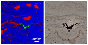
Image Raman ultrarapide SWIFT™ d'une coupe minérale, illustrant la distribution des variétés de quartz (rouge/bleu) et un filon d'hématite.
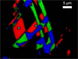
Image Raman SWIFT™ de graphène sur silicium, montrant des zones monocouches, bicouches et tricouches. Une taille de pas de 200 nm a été utilisée, avec une intégration de 50 ms par point.
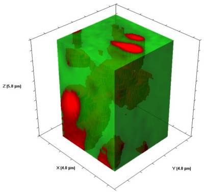
Affichage Raman 3D SWIFT™ de particules de sulfate de baryum dans une matrice de polymère, dans un volume de 92 µm3. Acquisition avec une taille de pas de 200 nm et une intégration de 50 ms.
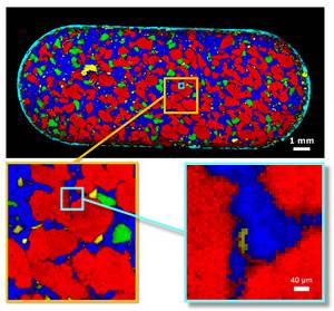
Image SWIFT™ haute définition d'un comprimé pharmaceutique entier de 17 mm, comprenant plus de 2,6 millions de spectres. Les parties zoomées sont extraites des mêmes données et montrent comment une seule image haute définition permet d'effectuer des relevés à l'échelle millimétrique et d'étudier les détails à l'échelle micrométrique.
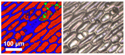
Imagerie Raman ultrarapide SWIFT™ de cellules d'oignon, illustrant la structure cellulaire générale (rouge/bleu) et les zones isolées d'espèces de caroténoïdes.
