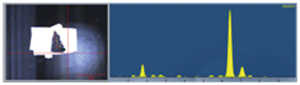
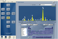
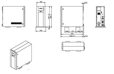
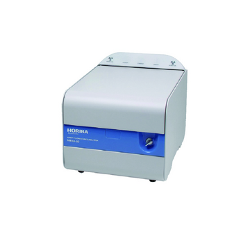
润滑油中磨损金属及添加剂元素分析

 您正在浏览的产品已停产,但您仍可以继续访问此页面以获取相关售后服务支持。
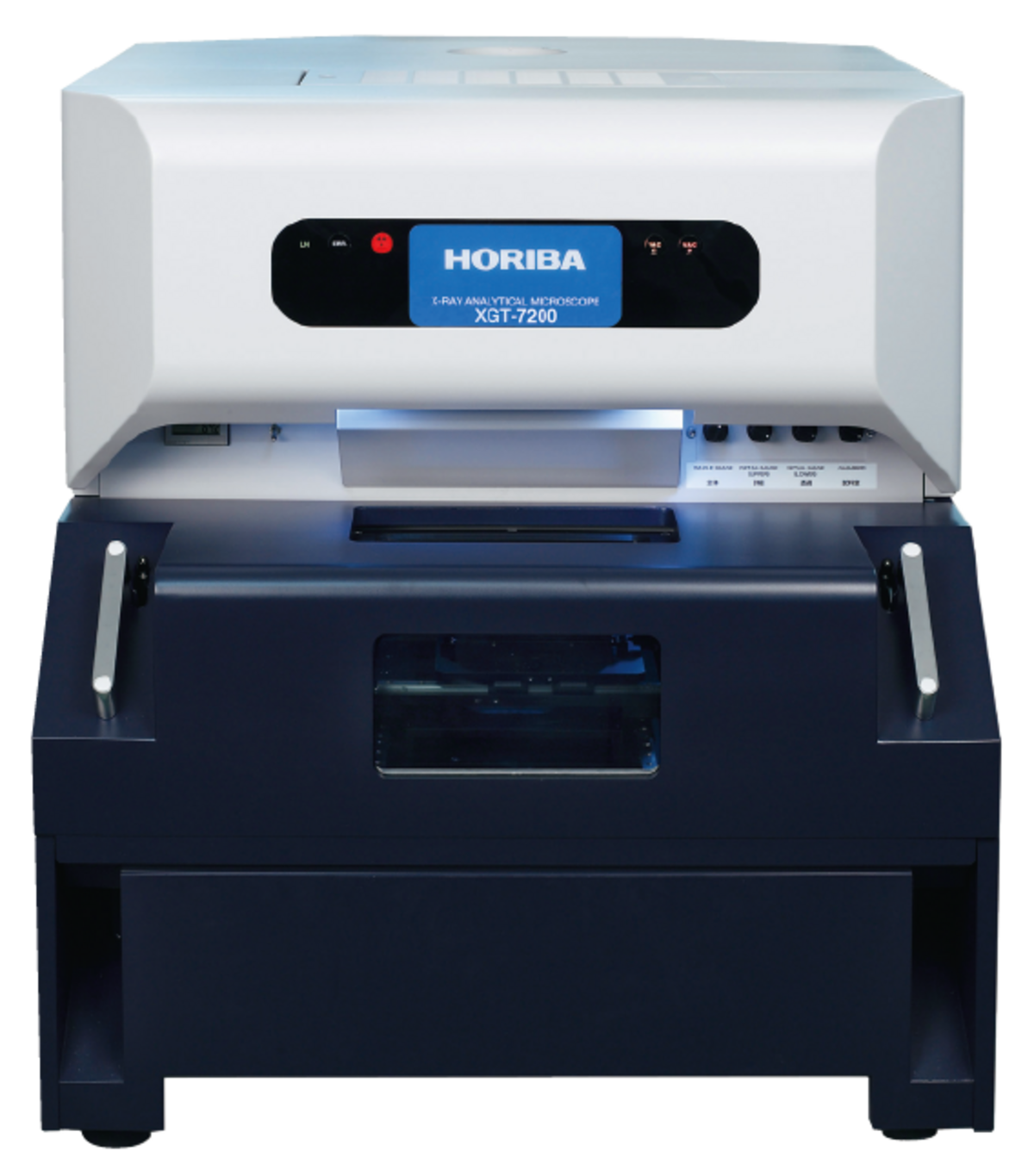
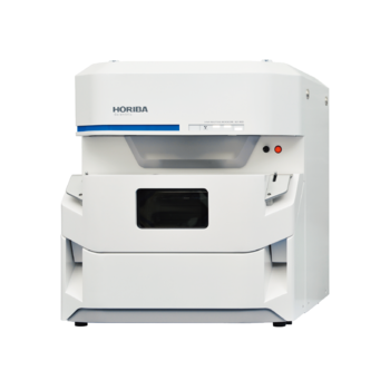
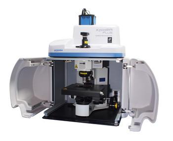
X-ray Analytical Microscope
The XGT-7200 represents a completely new generation of XRF microscope, and leads the way to a new era of science. It offers a seamless merger between optical observation and elemental analysis functions, revolutionizing the world of micro-analysis and establishing micro-XRF as a routine tool for the research and analytical scientist.
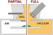
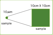
Unique hardware features ensure the system offers versatility and flexibility for every measurement. A choice of two software controlled x-ray guide tubes with diameters ranging from a unique 10 µm through to 1.2 mm allow conditions to be optimised for a range of measurements, including both micro and macro. Similarly, with the unique Dual Vacuum Modes it is possible to switch within seconds between a high sensitivity full vacuum mode and a versatile localised vacuum mode. The latter maintains samples at atmospheric pressure whilst retaining sensitivity to all elements from sodium to uranium.
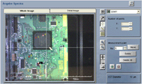
In any configuration the intelligent combination of optical cameras ensure that the precise analysis position can be quickly and simply located.
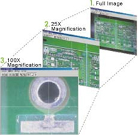
Quickly proceed from a view of the entire sample to the selection of the analysis potision.
Intuitive "click and move" images from low magnification and high magnification cameras within the sample chamber, allow the analysis position or mapping area to be defined withing seconds.
Single Point and Multi-point Analysis
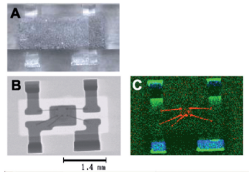
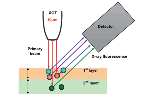
Single point and automated multi-point analyses allow high quality spectra to be acquired from either a single position, or from a number of user defined points across the sample. Element peaks are automatically located and labelled, and quantitative analysis down to ppm levels can be carried out using the fundamental parameters method (FPM), FPM with single standard, and full standard sample calibration.Thickness calculations can also be made on nm and µm thick multi-layered structures.
Hyperspectral Mapping Analysis
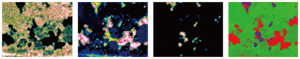
The SmartMap imaging software records a full EDXRF spectrum at each and every pixel of the element image, enabling post-acquisition element image generation and comparison, and spectrum generation from user defined regions in the image with subsequent qualitative and quantitative characterisation.
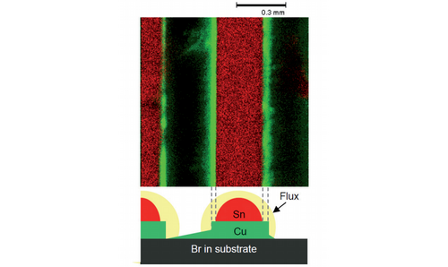
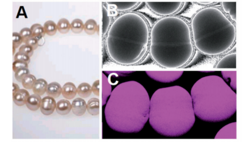
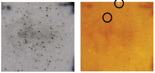
Transmitted X-ray imaging provides additional insight into a sample’s structure, allowing features invisible by eye to become immediately apparent.
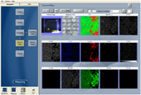
Element image display
Element images to be displayed can be selected during and after acquisition. Images can aslo be generated from user defined spectral windows.
RGB Composite Image Generation
Overlay elemental images to allow easy comparison of element distribution
Spectrum Generation
Generate the average spectrum from a user defined region within an image.
Line Analysis
Display multiple element intensity profiles across a defined region of an image.
Data Export
Element images can be exported in data or image formats. The entire hyperspectral datacube can be exported in RAW format.
The unique features of the XGT-7200 have seen this innovative micro-XRF analyser widely embraced for a range of applications, including electronics, engine wear analysis, forensic science, geology, mineralogy, pharmaceutics, museums, metallurgy, biology, medicine and archaeology.
The flexible XGT-7200 micro-XRF system covers everything from macro analysis, for a general survey of a wide area, to the inspection of a specific micro area, with simultaneous XRF and transmission imaging. Its many features ensure high performance analysis with easy operation.


XGT-7200 Specification
Elements: Na to U
X-ray tube: Rh target / Tube voltage 50kV / Tube current 1mA
Fluorescent X-ray detector: Peltier cooled Silicon Drift Detector (SDD)
Transmitted X-ray detector: Nai(Tl) scintilltor
X-ray guide tube: Mono capillary 10μm / 100μm with no filter
Optical image: Full sample optical image and coaxial magnification image
Sample stage size: XY : 100mm×100mm
Sample chamber: Vacuum chamber model/ Max 300mm×300mm×80mm in vacuum
Signal processing: Digital pulse processor ( INCA unit )
Qualitative analysis: Auto identification / KLM marker / Peak search / Compare spectrum
Quantitative analysis: Non standard FPM / Standard FPM / Standard file matching FPM /Calibration curve / Multi layer FPM (Thickness gage) /Multi point analysis (Max5000) / Multi point result send to Excel / XGT-5000 spectrum view
Mapping function: Transmitted X-ray image / Elemental image / Spectrum mapping / Rectangle mapping / Generate spectrum / RGB composition / Scale maker / Line analysis
Other function: Possible to start the XGT-5200 software simultaneously.

如您有任何疑问,请在此留下详细需求信息,我们将竭诚为您服务。
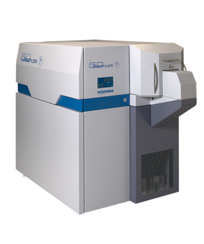
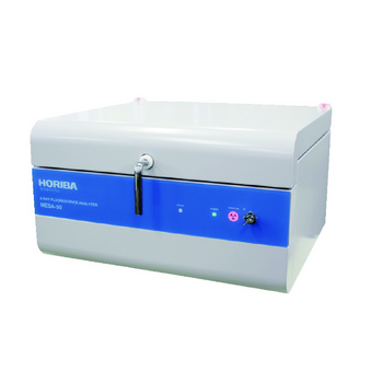
X 射线荧光分析仪
X射线荧光分析仪
高分辨率,高灵敏度和高稳定性的电感耦合等离子体发射光谱仪
高性价比的电感耦合等离子体发射光谱仪
微区X射线荧光分析仪
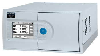
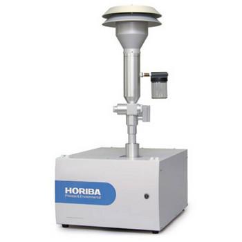
空气污染监测仪
空气颗粒物监测仪
空气颗粒物监测仪
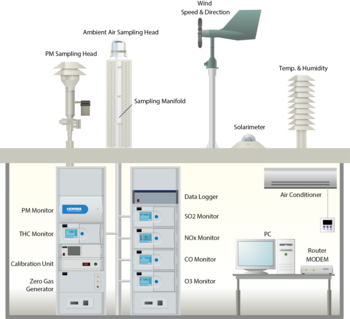
空气质量监测系统

一键式全自动快速椭偏仪
定制化专属设备

Auto-Soft用户导向的分析界面-用于Auto SE和Smart SE
化学和显微白光图像混合显示
功能齐全的椭圆偏振光谱软件
自动 DLC 涂层分析
优化的分析流程
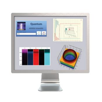
Quantum 和Image
适用于各种形状、尺寸和特性的样品附件
用辉光放电光谱仪去发现一个崭新的信息世界

ICP Software

Complete your ICP-OES Spectrometer for your specific needs
增强拉曼和光学图像

For your specific application
拉曼光谱搜索

纳米拉曼光谱仪

高分辨超灵敏智能拉曼成像仪

纳米拉曼光谱仪
LabSpec 6是经过验证的软件

基于实时激光干涉测量的摄像头终点监测
X 射线荧光分析仪
X射线荧光分析仪
调用设置,采集过程自动化

固体颗粒计数系统
多位置自动获取拉曼光谱
适用于所有拉曼成像的多变量分析应用程序

车载排放测量系统
快速简单的拉曼采集

高通量、开放式表面等离子体成像仪
测试、识别和分类粒子

符合人体工程学的软件,兼具数据采集,数据处理和技术支持
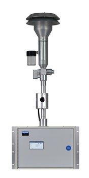
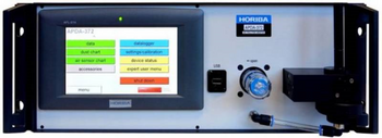
带 X 射线荧光的连续颗粒物监测仪
使用VBS进行定制化
自动硅应力分析

智能型多功能椭偏仪
将拉曼成像从几小时变成几分钟

高分辨率,高灵敏度和高稳定性的电感耦合等离子体发射光谱仪

高性价比的电感耦合等离子体发射光谱仪

真空紫外椭偏仪

在线监控椭偏仪

研究级经典型椭偏仪
微区X射线荧光分析仪
微区X射线荧光分析仪超大样品仓型

纳米拉曼光谱仪
高性能全自动拉曼光谱
复杂数据集的数据分析
密码保护的用户访问控制

快速、灵敏、高分辨的深度剖析技术
X 射线荧光分析仪
X射线荧光分析仪
X射线荧光油中硫/氯分析仪
带 X 射线荧光的连续颗粒物监测仪
X射线荧光油中硫分析仪
X射线荧光油中硫分析仪
微区X射线荧光分析仪
微区X射线荧光分析仪超大样品仓型