discontinued Ce produit a été abandonné et n'est plus disponible. Vous pouvez toujours accéder à cette page à des fins d'information.
successor
Les caractéristiques de ce produit ont été intégrées dans un nouveau modèle. Continuer vers le successeur

XploRA INV

Inverted Raman Microscope

The XploRA INV inverted Raman microscope combines the automation features and small footprint of the standard XploRA™ Raman microscope with the unique sampling capabilities of an inverted microscope, especially important for demanding biological applications.
 

Segment: Scientific
Fabricant: HORIBA France SAS

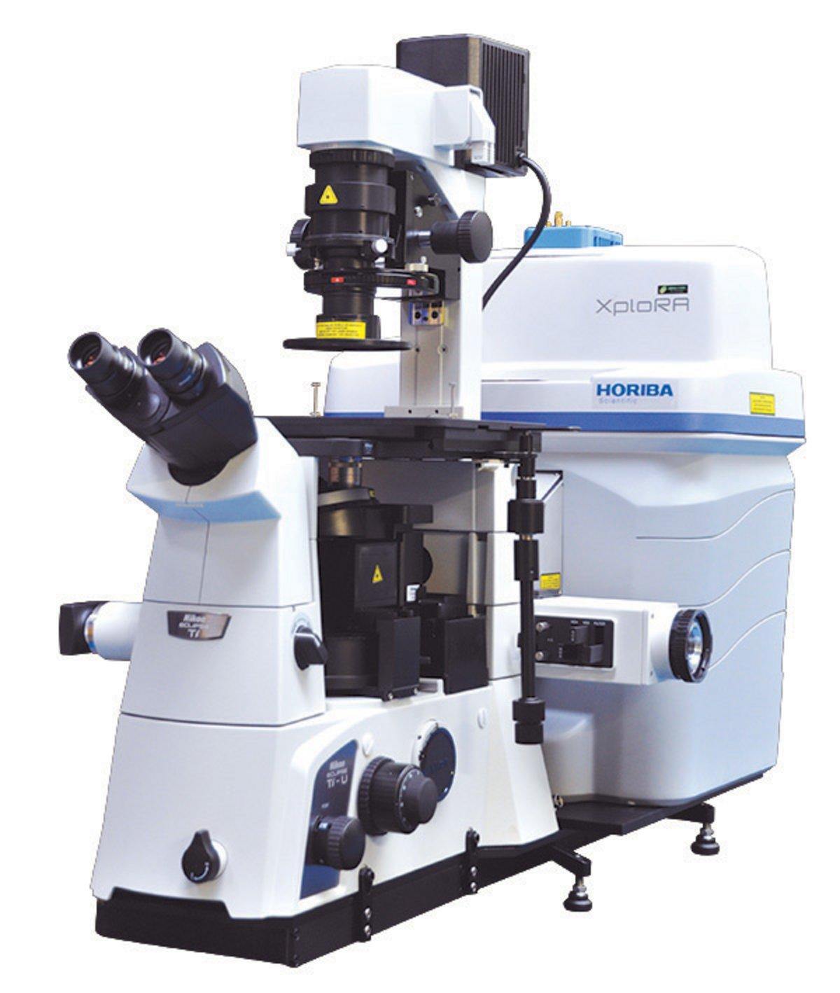
The XploRA INV combines the exclusive automation features and small footprint of the standard XploRA™ Raman microscope with the unique sampling capabilities of an inverted microscope, especially important for demanding biological applications. These applications include:

  • cell research
  • disease detection
  • characterisation of drug-cell interactions
  • pharmaceutical verification of intercellular activities


In addition, the inverted geometry allows easy incorporation of AFM units for combined Raman-AFM and TERS (Tip Enhanced Raman Spectroscopy) for highest spatial resolution sample visualisation.

The open structure of the XploRA INV also permits the use of virtually all options and add-ons for inverted microscopes, including micro manipulators and “optical tweezers”, specific enclosures for cell applications, etc.

The XploRA INV also offers the option to integrate the unique scanning capabilities of HORIBA’s patented DuoScan for rapid spectroscopic multichannel Raman imaging, as well as the new 3D FCI (Fast Confocal Imaging) module, which allows ultra-fast fluorescence imaging for visualisation and verification.


White light image (left), fluorescence FCI image (center) and hyperspectral fluorescence image (right) images of a bovine embryo after staining with Nile Red, acquired on the XploRA INV.  The hyperspectral image illustrates the stain in different polarity states.  Data courtesy of Prof. Igor Chourpa, Université de Tours, France.


Faster Raman                                                      YES with XY stage                                                                                                           
SWIFT™ Imaging                                         Optional DuoScan™ imaging           
/ SWIFTXS (with
EMCCD)


Confocal Imaging                                                 0.5 μm XY                                                                                                         


Routine operation                                              Methods and scripts 
Automation


Full Microscope                                                  Inverted


 Resolution                                                         Standard + High
                                                                          

 Multi-laser                                                       532, 638, 785 nm
  Options                                                          others on request
                                                                     

Assessing Biotherapeutics Stability using Raman Spectroscopy
We have measured solutions of lysozyme under conditions known to effect its physical state in order to investigate the potential of Raman spectroscopy as a non-invasive and label-free tool to assess protein formulation stability. Results from this study identified specific Raman signature bands in this protein that can be used to identify individual amino acid residues that are reflect structural changes in proteins.

Request for Information

Do you have any questions or requests? Use this form to contact our specialists.

* These fields are mandatory.

Related products

OpenPleX
OpenPleX

Manual label-free molecular interaction analysis machine Flexible Research Platform

Sanitary Conductivity Sensor (Ferrule Clamp type) FES-210L

Insertion sensor type for wide-range conductivity meter HE-960HI

XploRA™ PLUS
XploRA™ PLUS

Spectromètre Raman – Microscope confocal Raman

LabRAM Odyssey
LabRAM Odyssey

Imagerie Confocale Raman et Spectromètre à Haute Résolution

LabRAM Odyssey Semiconductor

Cartographie Raman et Photoluminescence de wafers

LabRAM Soleil
LabRAM Soleil

Microscope Confocal Multimodal Raman

MacroRAM™
MacroRAM™

Spectromètre Raman de paillasse abordable

Microscope Raman modulaire

Système Raman Polyvalent

nanoGPS navYX
nanoGPS navYX

Microscopie corrélative collaborative

Raman Spectrometer - MINI-CCT+

MINI-CCT+ Mini Raman Spectrometer

Sondes à fibre Raman

Spectromètres Raman

Sondes Raman miniatures OEM

Systèmes et composants Raman miniatures OEM

Spectrographe HE
Spectrographe HE

Spectromètre Raman haute efficacité dédié pour un suivi Raman des procédés

Spectromètre Raman térahertz modulaire

Spectromètre Raman abordable à ultra-basse fréquence, jusqu'à 10 cm-1

Spectromètre Raman UV

pour les spectroscopistes Raman UV

XploRA™ PLUS
XploRA™ PLUS

Spectromètre Raman – Microscope confocal Raman