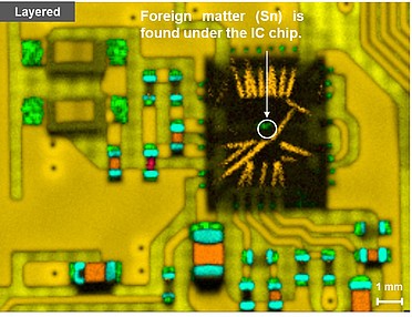
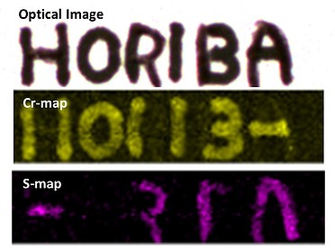
Spectrométrie de Fluorescence X
Biology / Medicine
Application Notes

- Bio01 (XGT01) : Biological applications of XRF microscopy.
- Bio02 (XGT02) : Elemental micro-analysis of leaves using EDXRF.
- Bio03 (XGT12) : Elemental analysis of single rice grains using XRF micro-analysis.
For a list of all available application notes please click here
Articles
- New Dimensions in Micro-XRF Elemental Analysis (G.I.T. Laboratory Journal, Issue 4, 2005)
- Spectroscopic Imaging for hte Life Sciences - More Than Just a Pretty Picture (Spectroscopy Europe Life Science Supplement, August/September, 2005)
- Advancing Towards a More Detailed Picture of Chemistry (European Biopharmaceutical Review, Spring, 2005)
For a list of all available articles please click here
Traditionally XRF has not been the must useful technique for biological and medical research, since it is usually necessary to work with a full vacuum in order to detect many of the biologically important elements (including Sodium, Magnesium, Sulfur , Calcium). Typical samples in these fields cannot withstand such experimental conditions.
However, the unique Partial Vacuum Mode available on the HORIBA XGT systems now opens this technique to the analysis of biological tissues and other water containing materials. Hence it is now possible to characterise these materials using XRF. The microscopic capabilities of the XGT systems allows individual particles or features to be analysed, and imaging experiments can show the distribution of elements across a surface.
Areas which have already utilised the power of micro-XRF analysis include bone disease research, drug pathways and accumulation in tissue, micro-nodules in plant leaves, phytoremediation pathways in plants, and imaging of gallstones. The common link between these is the requirement for microscopic elemental analysis of both major and trace elements.
Key areas which benefit from micro-XRF include:
- Leaves and plants
- Tissue
- Bones
- Disease investigation
- Drug interactions
- Pharmaceutical materials
- Mineral deposits and stones
- Implants and scaffolds
Example

Analysis of mulberry leaves illustrates an internal structure which varies with leaf age. The transmitted X-ray image shows the main tissue of the leaf with the transport vessels clearly visible. In the older leaf (C) Calcium nodules are clearly visible, whilst the youngest leaf (A) shows none. It is surmised that these nodules could be a defence established against silk worms, for whom the mulberry is a principal food stock.