Size:
0.73
MB
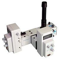
Microscope PMT Photometers Specification Sheet

Ideal for Electrophysiology researchers to quantitate light intensity out of a microscope
HORIBA's microscope photometers are ideal for any lab wishing to quantitate light intensity from a sample on a microscope stage. Initially designed for the most demanding low light level fluorescence kinetics of labeled mammalian cells, these photometers are also just as well suited for mineral analysis or transmission studies.
The photometer was developed by Photon Technology International (PTI) with over 1,000 installed systems around the world. PTI is the pioneer in the quantitative ratio fluorescence marketplace having introduced the first patented ratio fluorescence system, the Deltascan™, shortly after the first Fura-2 publication.
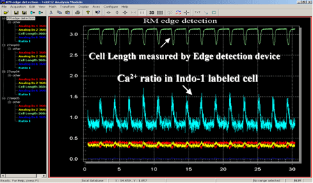
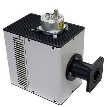
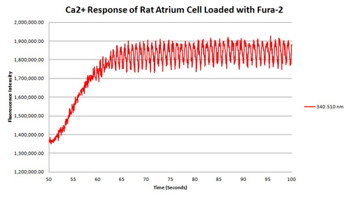
Dual emission channel photometer for Indo-1 Ca++ measurements
The above data traces are from the simultaneous collection of fluorescence and cell length from a cardiac myocyte. Myocyte was loaded with dual emission ratiometric fluorescence probe Indo-1. The DeltaRAM illuminator was used for excitation illumination at 365 nm and the emitted fluorescence was detected with a dual emission PMT photometer. Above data was collected with FeliX32 photometry software from Photon Technology International (PTI). The blue trace shows the calcium ratio increase and decrease with the cell contraction. The contraction data (Green trace) was detected with a video edge detection electronics module and is correlated with the fluorescence data since both signals were collect simultaneously.
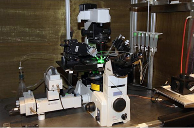
Image courtesy of, Prof. Dr. György Panyi, Department of Biophysics and Cell Biology, University of Debrecen
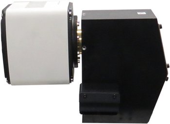
Shown is a picture of a PTI/OBB dual emission photometer attached to the C-mount of an inverted fluorescence microscope. The microscope is also equipped with electrophysiology recording devices and perfusion apparatus. The entire setup is inside a Faraday cage for electrical isolation.
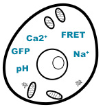
HORIBA's photometers are ideal for quantitative live cell measurements of intracellular ion and molecules such as Ca++, Na++, pH, GFP, FRET and FRAP experiments. By allowing for simultaneous quantitative fluorescence detection with patch clamp recordings, these photometers have been widely used by electrophysiologists around the world. The PMT photometer is a passive detection system that provides an output voltage signal proportional to the ion/fluorophore of interest, and this signal is directly fed into the electrophysiology A/D converter and collected with the customer’s existing software. The PMT photometer can collect a fluorescence signal at up to 20 KHz for high speed transient recordings.
While the PMT photometer was initially designed for demanding low light level fluorescence detection, it is extremely well suited for a variety of other microscopy applications including the following.
The standard viewing eyepiece can be replaced with an optional backlighting LED to allow the user to set regions of interests without ever needing to move from the microscopes eyepiece.
With this option the LED then illuminates the field of view through the adjustable iris. You can then look at the field of view through the microscope eyepieces and adjust the LED lit square or rectangle with the edges of the targeting iris.
For some applications like single molecule detection you may need to detect from precisely known physical areas in the field of view. For such applications having a variable iris is not sufficient, so there is an optional pinhole insert that replaces the adjustable iris
The pinhole assembly is a mechanical insert that goes in place of our standard aperture blade assembly. There is a one-inch diameter holder for laser cut pinhole apertures with holes that can be selected from 5 µm to 200 µm in diameter (select from list). The pinholes are positioned in the focal plane of the microscope and can be translated +/- 3 mm from center with easy to use micrometers.
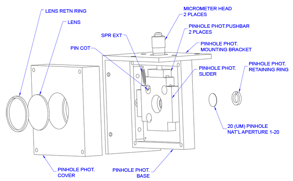
Since the pinholes are so small in diameter, the only way to target through them is by backlighting the pinhole with our backlit LED option and visually aligning them in the field of view through the eyepieces of the microscope. The user can target a specific region of interest for the photometer to measure by using the X/Y translation on the pinhole insert or by moving the stage of the microscope.
The multi channel microscope photometers are designed to hold standard 18mm dichroic cubes, with 18 mm circular bandpass filters and an 18 x 26 mm dichroic mirror. Currently we offer three standard application specific dual emission cube assemblies that include two emission filters and an appropriate dichroic mirror. Custom filters and dichroics may be purchased separately.
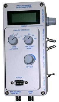
All controls and displays are available on the front and side of the PMT housing.
Detector Control Specifications | ||
|---|---|---|
| Display | LCD display of high voltage or signal |
Controls | On/Off switch | |
BNC Connectors | Signal Out | |
| Input | ± 15 VDC, 250 mA |
|---|---|
| High Voltage | -200 to -1,100 VDC manually adjustable LCD displays actual cathode voltage |
| External High | 0 to +5 VDC (0 = -200 V, 5 = -1,100 V) |
| Voltage Adjust | Continuously adjustable |
| Input Regulation | ± 0.05% max. (for 15 V ± 1 V input). |
| Load Regulation | ± 0.05% max. |
| Ripple | 100 mV p-p max. |
| Temperature Coefficient | ± 0.01% max. (+5 to 40°C) |
| Drift | ± 0.03%/hr. max. (after 15 minute warm-up) |
| Input | 115 or 220 VCA (specify at time of order) |
|---|---|
| Output | ±15 VDC, 250 mA |
| Gain Settings: | 1 µA = 1 V 0.1 µA = 1 V 0.01 µA = 1 V 0.001 µA = 1 V |
|---|---|
| Time Constant Settings: (with Corresponding Analog Frequency) | 0.05 msec (20 KHz) 0.5 msec (2 KHz) 5 msec (200 Hz) 50 msec (20 Hz) 500 msec (2 Hz) |
| Offset Correction: | ± 50 nA |
| Signal Output on BNC connector: | Analog voltage |
| Linear Dynamic Range: | 5 orders of magnitude* |
|---|---|
| Dead Time: | 250 ns |
| Signal Output on BNC connector: | TTL |
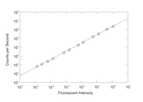
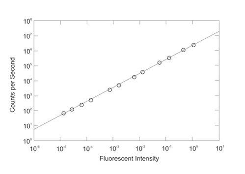
* Linear dynamic range of the photon counting PMT housing. The intensities were produced by attenuating the fluorescence emission of a fluorescein sample with neutral density filters.
| Dimensions (WxDxH) | 4 x 4 x 8 inches |
|---|---|
| Weight | 4 lbs |
Do you have any questions or requests? Use this form to contact our specialists.
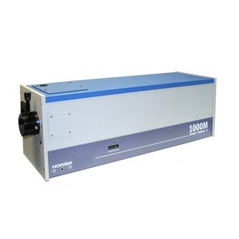
Long Focal Length Spectrometer
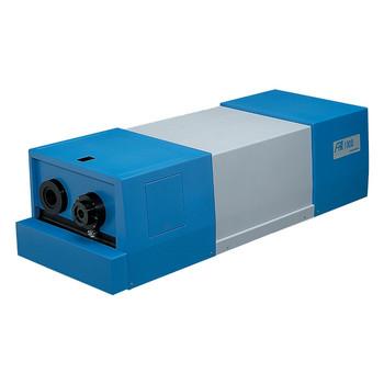
Dual Mode Analog/Photon Counting PMT
High Resolution Monochromators
Double monochromator
Mid-Focal Length Imaging Spectrometers
Short Focal Length Imaging Spectrometers
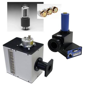
Self contained PMT housing for quantitative spectroscopy and imaging low light measurements
Deep Cooled UV/Vis/NIR
Large choice of PMTs, solid state, photoelectric detectors for custom spectroscopy solutions
EMCCD Scientific Camera
Deep Cooled NIR Scientific Cameras
OEM CCD Camera
Deep Cooled Vacuum Ultra Violet Scientific Cameras