Discover what our customers are saying about HORIBA’s Raman Spectroscopy solutions! From authentic testimonials to hands-on success stories, their voices highlight how our instruments empower real-world research and innovation. In the Science in Action Series, you’ll find inspiring examples where Raman technology helps solve practical challenges, advance scientific understanding, and open new possibilities across a wide range of applications.
![]() University of La SAPIENZA, Rome, Italy - Claudia Fasolato, Research Scientist:
University of La SAPIENZA, Rome, Italy - Claudia Fasolato, Research Scientist:
The LabRAM Soleil is so compact, well-illuminated and versatile that you can measure any kind of sample with it!
It’s the ideal tool for our physics and biophysics research group, where we’re working on a wide variety of applications from nano-objects to perovskites and biological samples. ![]()
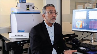
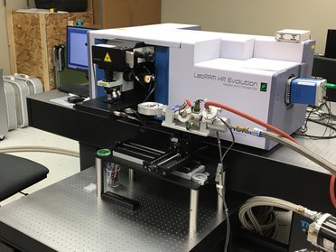
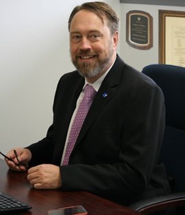
![]() VIBRA-SANTÉ HUB, Center for Interdisciplinary Research on Medicines, Liege, Belgium – Eric Ziemons, Director:
VIBRA-SANTÉ HUB, Center for Interdisciplinary Research on Medicines, Liege, Belgium – Eric Ziemons, Director:
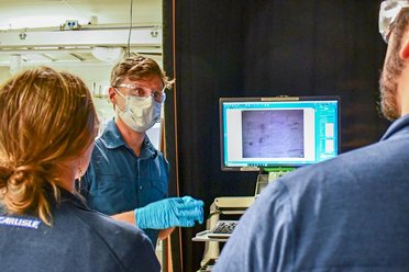
With a multimodal Raman microscope as easy to set up as the LabRAM Soleil, we were quickly up to speed. We were able to study our pharmaceutical samples safely and without risk of contamination as it has Class 1 capability built in. It’s the ideal tool for the analyst of the future! ![]()
![]() IFREMER, Brest, France – Maria El Rakwe, Research scientist:
IFREMER, Brest, France – Maria El Rakwe, Research scientist:
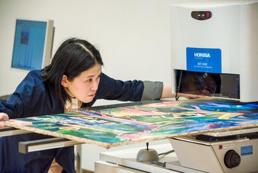
We really like the LabRAM Soleil’s optimized design, which speeds up analysis of microplastics. It has helped us to improve our profitability, no matter what kind of environmental sample we’re looking at (notably water). ![]()


Do you have any questions or requests? Use this form to contact our specialists.