PDF
0.47
MB

Heme Insights 8_August 2026_HORIBA
Discover how to investigate unexpected low platelet counts with a practical case study, diagnostic pathway, quiz, and laboratory best practices.

When an apparently severe thrombocytopenia is caused by platelet aggregation rather than a true fall in circulating platelets
A 69-year-old male had a platelet count of 41 x 10^9/L and hemoglobin of 9.0 g/dL. The automated result appeared to indicate severe thrombocytopenia.
| WBC 15.1 x 10⁹/L | MCV 90.0 fL |
| RBC 2.85 x 10¹²/L | MCH 31.6 pg |
| HGB 90 g/L | MCHC 350 g/L |
| HCT 0.257 L/L | PLT 41 x 109/L |
| Patient: Male, 69 years |
| Platelets: 41 x 10^9/L |
| Blood smear: Extensive platelet aggregation |
| Interpretation: Pseudothrombocytopenia |
| Action: Check for clots and repeat in citrate |
Differential: neutrophils 76.1%, lymphocytes 7.1%, monocytes 5.1%, eosinophils 0.0% and basophils 0.0%.
On manual inspection, extensive platelet aggregation was present. The apparent low platelet count was therefore interpreted as false thrombocytopenia caused by platelet clumping. Large aggregates may also interfere with other analyzer measurements.
Recommended action
Inspect the specimen for a clot and request a correctly filled citrate sample for prompt repeat analysis, following the laboratory's validated procedure.
Example of Cells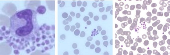
Why can the platelet count be falsely low?
When platelets form aggregates, an automated analyzer may exclude the clump, classify it incorrectly or generate a platelet-clump flag. The reported count can therefore be substantially lower than the true circulating count. This in-vitro phenomenon is known as pseudothrombocytopenia.
Key Message: Never accept an unexpected low platelet count without checking the sample, analyzer information, and a blood smear.
A structured approach helps distinguish a pre-analytical or EDTA-related problem from genuine thrombocytopenia
1. Start with the sample
Inspect the tube for visible clotting and assess sample quality. Difficult venipuncture, slow collection, underfilling or inadequate mixing can allow platelet and fibrin aggregates to form. A clotted or partially clotted sample is unsuitable for an accurate platelet count.
2. Review the analyzer
Check platelet-clump flags, histograms, scatter information and previous results. Consider whether the count agrees with the clinical presentation and whether large aggregates could also affect the white cell result.
3. Examine the blood smear
Review the full smear, including the feathered edge and lateral margins. Assess for platelet clumps, giant platelets, satellitism, fibrin strands, schistocytes, blasts and other significant abnormalities.
4. Repeat appropriately
When EDTA-dependent clumping is suspected, request a correctly filled citrate sample and analyze it promptly according to the laboratory’s validated procedure. Confirm microscopically that the clumping has resolved before releasing a numerical result.
EDTA-dependent pseudothrombocytopenia EDTA may expose platelet membrane epitopes which allow antibody-mediated aggregation in-vitro. This occurs in the sample tube and does not necessarily reflect the circulating platelet count. |
Reporting the result
Do not report severe thrombocytopenia as genuine when significant platelet clumping makes the result unreliable. Genuine thrombocytopenia and aggregation can occasionally coexist, so interpretation should include the platelet estimate, previous results, clinical context and repeat testing.
Suggested laboratory comment
“Platelet aggregates are present. The automated platelet count may be falsely reduced. Please send an appropriately collected citrate sample for confirmation, in accordance with local laboratory procedure.”
Learning points
| 1 Check the sample | 2 Review analyzer flags | 3 Examine blood smear | 4 Repeat in citrate when indicated |
A patient’s EDTA sample gives a platelet count of 41 × 10⁹/L. The analyzer flags possible platelet clumps, and the blood smear shows aggregates. What is the most appropriate next step?
a) Report severe thrombocytopenia immediately.
b) Request a correctly collected citrate sample and analyze it promptly.
c) Issue a normal platelet result based on the smear estimate.
d) Ignore the clumps and repeat the EDTA count the next day.
Answers are revealed two weeks after launch—check your inbox, our socials (LinkedIn / Facebook / X), or the next issue.
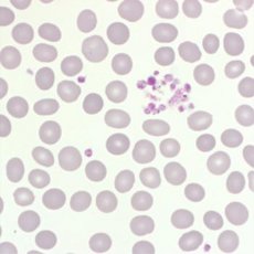
A patient presents with low Hb, RBC, normal MCV, but with very low platelet count. The blood smear shows the following red cell abnormality and occasional nucleated red cells. What condition may this be caused by?
a) Microangiopathic Thrombocytopenia in conjunction with Microangiopathic Hemolytic Anemia
b) Iron deficiency
c) Megaloblastic anemia
The Answer: a)
Numerous schistocytes and keratocytes are present, caused by mechanical fragmentation of red cells as they pass through partially occluded microvessels. The low platelet count occurs due to platelet consumption as they form microvascular thrombi.