Know What Your Particles Are—Not Just That They Exist
A particle count tells you something is present—but not what it is, where it came from, or whether it actually matters. That missing information slows investigations, creates uncertainty, and increases risk—particularly when particle size, shape, and composition all play a role.
Particle Correlated Raman Spectroscopy (PCRS) transforms particle analysis by combining automated microscopy‑based particle detection and morphological analysis with Raman‑based chemical identification in a single, correlated workflow.
PCRS delivers two critical dimensions of particle intelligence:
Together, these provide true single‑particle characterization, rather than isolated metrics.
Unlike traditional approaches that separate particle sizing from chemical analysis, PCRS enables a bidirectional workflow that adapts to your application needs.
For many applications, microscopy and image analysis guide Raman measurements:
Once particles are chemically identified by Raman spectroscopy, PCRS enables the workflow to move in the opposite direction:
This enables chemically resolved particle sizing and shape analysis, not just bulk averages.
By correlating Raman spectroscopy with particle imaging, PCRS enables:
With PCRS, particle analysis becomes actionable insight. You move beyond knowing that particles exist to understanding what they are, how they behave, and why they matter—supported by real, defensible data.
Proper sample preparation is critical for reliable PCRS analysis, as particle detection and Raman spectral quality depend on dispersion, substrate background, and optical access. Preparation workflows are designed to preserve particle integrity, minimize spectral interference, and ensure sufficient particle separation for automated analysis across applications including pharmaceuticals, batteries, carbon materials, forensics, and microplastics.
Dry particulates
Dry powders are lightly dispersed onto Raman-compatible substrates at low surface density to minimize overlap. Gentle tapping or use of the HORIBA XD-100 disperser helps reduce aggregation without damaging particles.
Liquid samples
Liquid samples are prepared by droplet deposition and drying or by filtration using Raman-compatible filters (e.g. silicon filters) and a standard filtration kit. Filtration conditions are optimized to capture representative particles while avoiding stacking or clogging. Simple liquid formulations such as nasal sprays may be prepared by spraying directly onto a slide and allowing the sample to dry prior to analysis. Special sample holders allow for automatic measurement of up to three silicon filters at once.
Standard filtration kit
Sample Holders
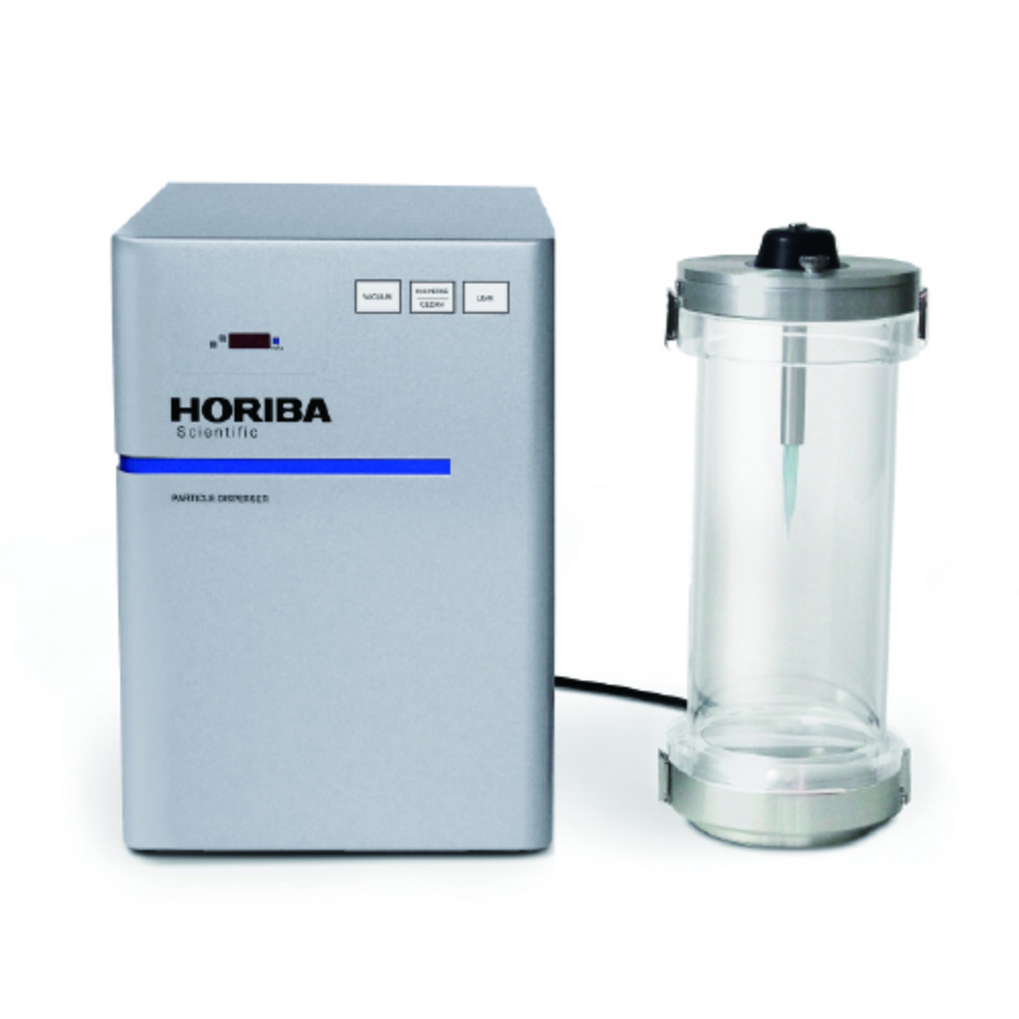
XD-100 Particle Disperser
Semi‑solid and viscous matrices
Gels, creams, and other viscous materials are applied as thin layers or diluted before filtration. Thickness is controlled to keep particles optically accessible while minimizing background interference.
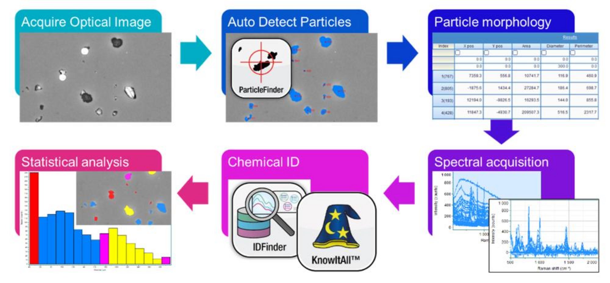
High-resolution optical images of the particles are acquired using various imaging modes such as bright field, dark field, transmission, or polarized light microscopy. Mosaic images may be acquired to cover all particles within a larger surface area.
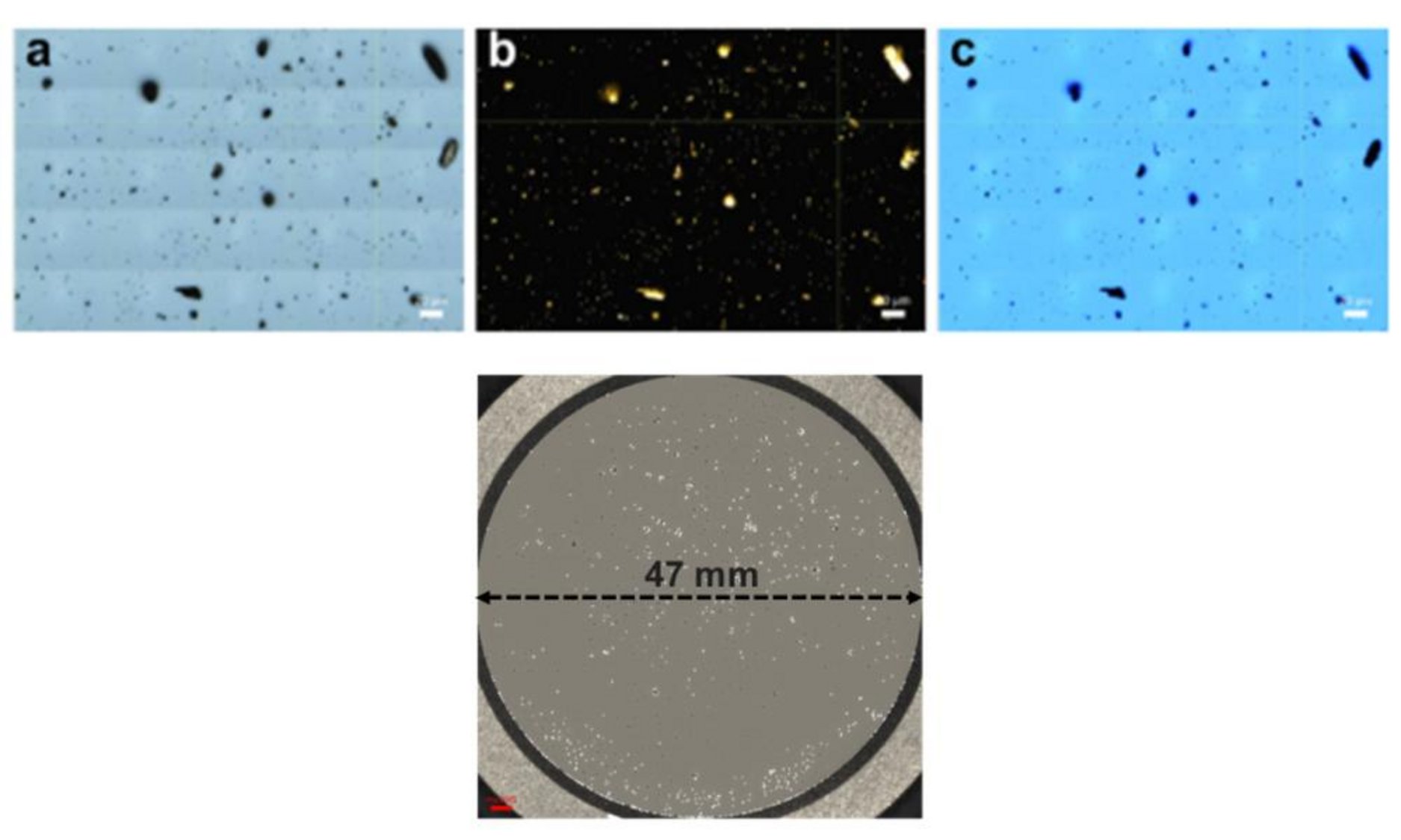
Particles are automatically detected according to various thresholding algorithms. Automated detection reduces operator-dependent variability and facilitates consistent screening of large particle sets.
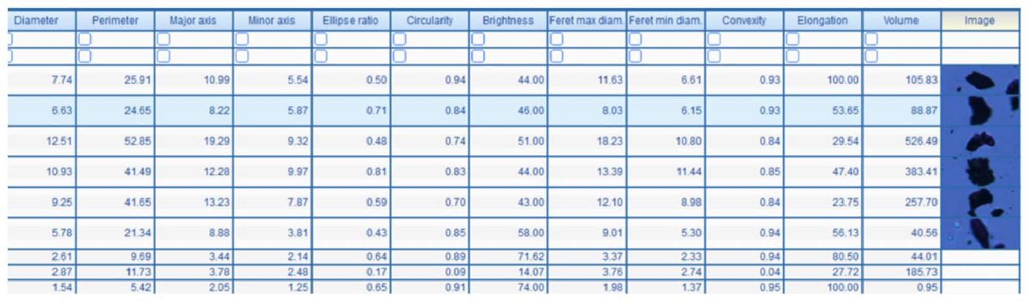
The ParticleFinderTM application automatically calculates size, shape, and morphological parameters for each particle, as well as their statistics. Users may filter particles to those most relevant or include all in the statistics. Get information on position, area, diameter, perimeter, major/minor axis, ellipse ratio, circularity, brightness, Feret max/min diameter, convexity, elongation, and volume.
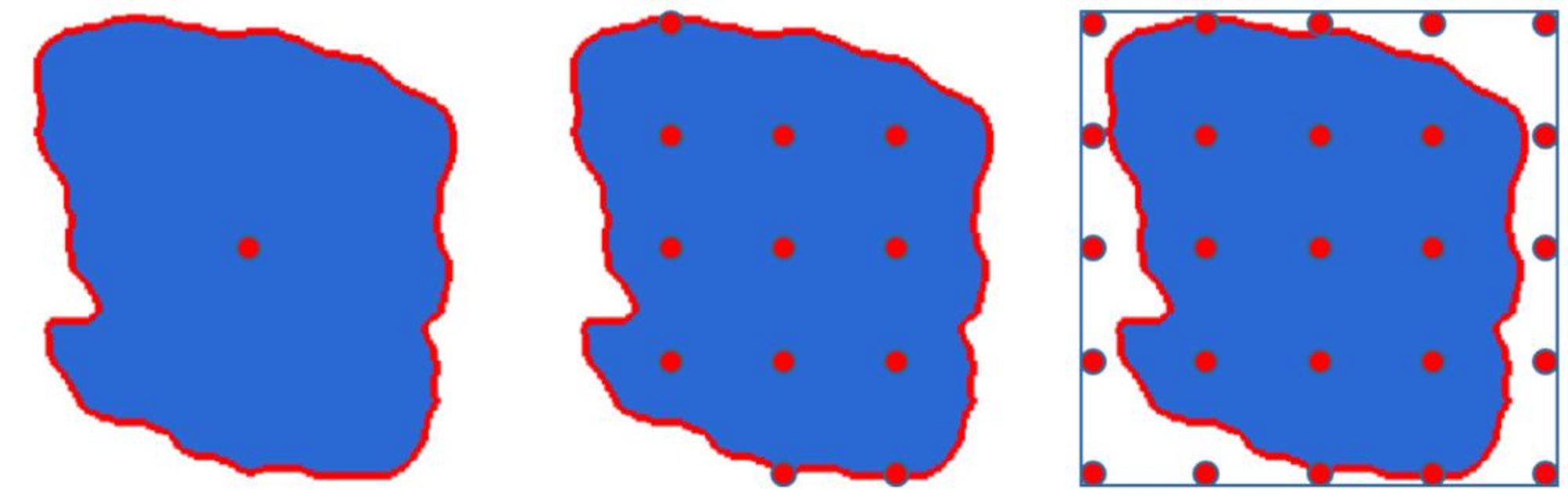
Raman parameters such as laser power, grating, integration time, etc. are optimized for the sample and Raman spectra are collected. Spectra may be collected using single point measurements at the center of the particle, multi-point averaging for heterogeneous particles, or full-particle level mapping, when necessary. Raman spectra are also processed (smoothing, baseline, spike removal, etc.) with the ParticleFinderTM application.
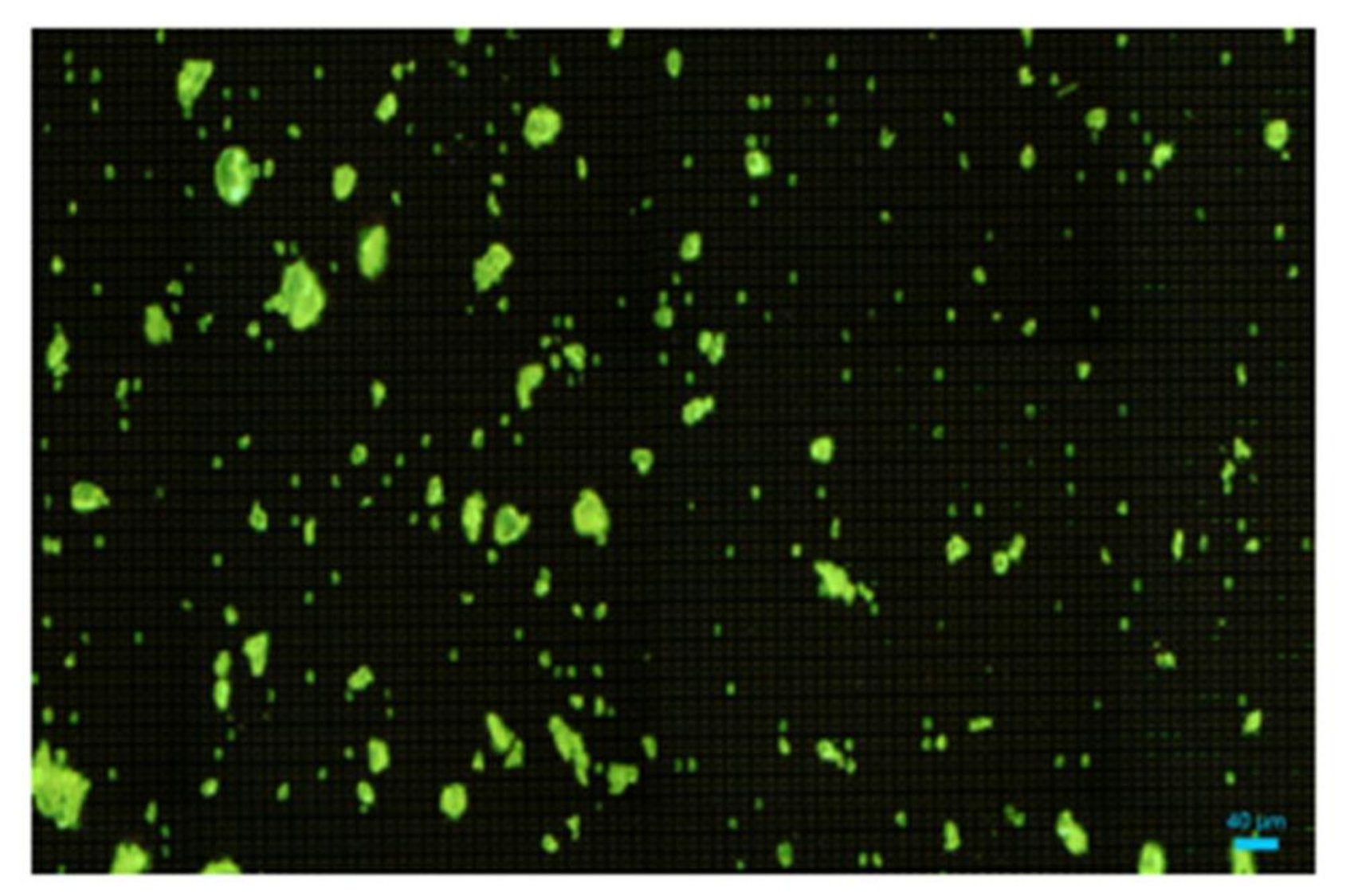
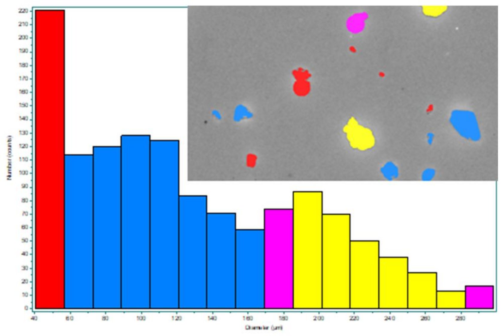
Each Raman spectrum is matched against a database, such as Wiley’s KnowItAll or HORIBA’s IDFinder in order to chemically identify each particle and color-code the particles according to material type.
![]()
![]()
All results are combined into a single dataset linking particle size, shape, morphology, and composition. A further statistical analysis can be performed as a function of particle ID and any of the relevant size, shape, or morphological parameters (e.g. ellipse ratio of all particles vs. ellipse ratio for particles of each type).
Particle Identification • Contamination • Raw Material Verification
PCRS supports pharmaceutical quality and forensic workflows by enabling:
✅ Supports pharmacopeial Raman approaches (e.g., USP <788>)
✅ Valuable for deviations, investigations, and QA/QC escalation
Environmental Monitoring • Particle Classification
PCRS is widely used for:
✅ Enables rapid screening and chemical confirmation
✅ Supports environmental research and monitoring initiatives
Contamination • Coatings • Materials Characterization
PCRS plays a critical role in advanced energy materials analysis, including:
✅ High spatial resolution supports localized defect analysis
✅ Useful across R&D, scale‑up, and failure investigations
Unknowns • Trace Evidence • Contamination
PCRS is trusted for forensic analysis due to its:
✅ Preserves sample integrity
✅ Supports investigative and evidentiary workflows
Particle Correlated Raman Spectroscopy (PCRS) is an advanced analytical technique that integrates:
Each particle is individually analyzed and fully characterized across Size, Shape, Location, and Chemical Composition. This creates a correlated dataset, delivering a complete understanding of particulate matter in complex samples.
PCRS is used for particle identification, contamination analysis, microplastics research, and failure analysis across multiple industries. Critical for contamination control, root cause analysis, and quality assurance.
Yes. Raman spectroscopy provides a molecular fingerprint that allows precise identification of unknown particles, including polymers, glass, metals, and biological materials.
PCRS enables rapid particle identification, correlated data analysis, automated workflows, and improved confidence in contamination investigations.
Traditional particle analysis methods answer:
PCRS answers the questions that actually drive decisions:
It transforms particle analysis from a reactive process into a proactive, insight-driven strategy.
MicroRaman Spectrometer - Confocal Raman Microscope

Raman Spectroscope - Automated Imaging Microscope

Confocal Raman & High-Resolution Spectrometer

Particle Disperser

Laser Scattering Particle Size Distribution Analyzer

Simultaneous Multispectral Nanoparticle Tracking Analysis (NTA)
Automated Particle Measurement, Identification and Classification using Raman Analysis
Spectral Identification Assistant within Raman Spectra Database
Raman Spectral Searching

Navigate your sharp image in real-time
Acquire the Topography of the Analyzed Sample & sharp, In-Focus Raman Images
Do you have any questions or requests? Use this form to contact our specialists.