
One of the greatest challenges in veterinary practice is faced when patients present with subtle or indistinct symptoms. At a glance, it can be hard to tell whether what may seem to be a slight niggling issue might in fact be an indication of something more serious.
More than most common pet ailments, inflammatory diseases can be especially tough to identify, often manifesting in low-key grumbling symptoms. When inflammatory conditions present in this fashion, they can easily go unchecked, bringing significant consequences for the health and overall quality of life of the patient.
To help combat the difficulties faced by veterinary professionals in identifying inflammatory disease, HORIBA UK Veterinary recently invited RCVS recognised specialist in small animal medicine, Dr Kit Sturgess FRCVS (Figure 1), to share his expertise on the matter as part of its successful CPD webinar series. The webinar, titled “Diagnosis of inflammatory disease in cats and dogs – Getting the most from your practice laboratory” included some valuable tips and insights on how best to rapidly assess disease severity in cats and dogs via inflammatory markers.
In this article, we will cover some of the key highlights from the webinar and provide practical advice including in-house diagnostic techniques to help support clinical decision making rapidly and cost effectively when faced with the sometimes vague presentation of inflammatory disease.

Figure 1. Dr Kit Sturgess RCVS, who presented on ‘Diagnosis of inflammatory disease in cats and dogs – Getting the most from your practice laboratory?’
Inflammatory disease encompasses a broad range of heterogenous conditions characterised by inflammation; the body’s response to injury, infection or irritation. Inflammation is a complex biological response involving immune cells, blood vessels and molecular mediators including cytokines and peptides. It is a protective mechanism that seeks to eliminate the initial cause of cell injury, clear out necrotic or damaged cells, and initiate tissue repair. The presence of inflammation therefore indicates a problem within the affected tissue, and usually requires treatment.
Inflammation can present in a variety of ways, with typical signs of redness, heat, swelling, pain and loss of function. While these signs might appear more evident when inflammation occurs locally or near the surface e.g., a painful, swollen joint, when inflammatory disease affects internal systems or organs, these tell-tale characteristics can be harder to spot.
Inflammatory diseases can manifest in distinct ways between individuals, and to differing levels of severity. Pancreatitis, a common inflammatory disease among cats and dogs, can range from severe acute cases with obvious outward symptoms, to chronic grumbling presentations which could have a good prognosis if caught early enough. The less severe, chronic condition accounts for the overwhelming majority of cases, highlighting the importance of making inflammatory disease detection easier.
Inflammation isn’t always isolated to a single organ or tissue. In some cases, it can instead present as a systemic condition. As with local inflammatory issues, systemic inflammation can either be acute, which is ordinarily a serious life-threatening condition, or chronic, which is less severe, and accounts for the majority of cases.
The more common chronic systemic inflammation can onset over a period of days or months, and occurs as a result of the release of pro-inflammatory cytokines from immune-related cells, and the subsequent activation of the innate immune system. In most cases, systemic inflammation is activated as a response to infection, neoplasia, or an immune-mediated disease. Recognising systemic inflammation is therefore a critical first step towards identifying these underlying conditions, but this form of inflammation often manifests in more subtle symptoms, like lethargy, mild appetite loss, and lower energy.
As with many diagnostic techniques in human medicine, biomarkers can be hugely valuable when assessing a veterinary patient with suspected systemic inflammation. A useful starting point is to investigate levels of acute phase proteins (APPs); blood-bound proteins that can be used to assess the innate immune system’s systemic response during inflammation. Serum-APP concentrations increase significantly in response to pro-inflammatory factors stimulated by the underlying disease process, and can be considered quantitative indicators of inflammation.
APPs have been monitored as biomarkers of inflammation for many years in the context of human medicine, but veterinary medicine has been slower to adopt this practice. APPs can be harnessed for diagnostic and prognostic purposes, as well as monitoring responses to therapy and for general health screening, and many of which can be readily tested for in-house.
Specific APPs demonstrate major, moderate or minor responses in times of inflammation, with stark differences in APP responses found between species. Major APP responders are found in low concentrations in healthy individuals, but rise 100 to 1000-fold during inflammation, making them ideal biomarkers of an inflammatory response.
The differing APP responses between species means that from a diagnostic point of view it is favourable to monitor levels of distinct biomarkers depending upon the species of the patient in question. For example, C-reactive protein (CRP) is a major-responding APP in canines, with serum concentrations increasing rapidly from <1 mg/L to >100 mg/L in times of inflammation. In cats however, CRP is not a major-response APP, and is therefore less useful.
Canine CRP can be highly useful in numerous conditions as its levels increase rapidly during systemic inflammation, rising within a few hours in response to pro-inflammatory cytokines, such as IL-6. Once inflammation has been resolved, CRP levels normalise within 36-48 hours, making for a highly sensitive biomarker of inflammation. Typically measured in serum via ELISA or immunoturbidimetric methods, CRP can be quantified against reference ranges relating to different inflammatory conditions.
As with any single parameter, CRP does have some limitations. Primarily, CRP is nonspecific, meaning that it cannot inform the underlying cause of inflammation i.e., infection, trauma or neoplasia. Furthermore, CRP only elevates when inflammation is systemic, making it unreliable for more localised inflammatory conditions. CRP therefore, can be useful for triage purposes in cases where a patient presents as generally unwell, or to detect systemic disease in patients with known localised inflammation.
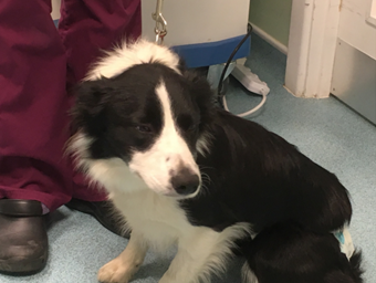
Figure 2. Munchy’s case study provided an excellent example of the value of monitoring CRP throughout the patient journey
In HORIBA’s recent webinar, Kit Sturgess talked through several canine cases in which CRP was vital for case management, diagnosis and monitoring responses to treatment. The interesting case of Munchy, the 13-month-old border collie (Figure 2) was a great example of the value of monitoring CRP throughout the patient journey. Munchy experienced a 4-day progressive history of gastrointestinal symptoms, lethargy and inappetence, and was referred by her first opinion practice for further investigation.
Munchy’s haematology report was inconclusive – with red and white blood cell counts within normal levels, while the biochemistry showed no elevated parameters to cause concern. Munchy was clearly unwell though, showing obvious signs of fever and discomfort. To investigate further, the veterinary team tested for CRP, which was found to be markedly elevated at 136 mg/L, suggestive of systemic inflammation. This result alerted the team that action should be taken and abdominal ultrasounds soon followed.
Scans showed the presence of granular abdominal fluid. Subsequent abdominocentesis revealed the presence of large numbers of degenerate neutrophils and a mixed bacterial population, along with some distension of her uterus, confirming septic peritonitis. For a patient with a severe infection like Munchy, one might expect the white blood cell count to be significantly elevated, but this example demonstrates the importance or conducting a thorough investigation. While leukocyte production increases during infection, many are sequestered into the abdomen, giving a haematology profile reading within the normal range.
Munchy underwent surgery for a hysterectomy and abdominal washout, and the team once again looked to CRP to monitor her progress. Day 1 post surgery Munchy was quiet and uncomfortable, with CRP elevated at 146 mg/L, but after two days she was brighter, more active and eating – with CRP down to 55 mg/L. Munchy went on to make a full recovery and was soon back to her favourite hobby, agility training!
The case study of Munchy, along with a range of other examples from the webinar, puts forward a strong rationale for monitoring CRP in suspected inflammation cases in canines. While it cannot be a comprehensive diagnostic tool, it is a reliable and sensitive measure that can direct veterinary case management, and indicate the speed and level of investigation required.
While CRP is a strong marker of systemic inflammation in canines, it is not an appropriate marker in feline subjects. At present, our understanding of cat APP responses is poor relative to canines, however some notable inflammatory markers have been identified. The APP most rapidly responsive to inflammatory and infectious conditions in cats is serum amyloid A (SAA), however more moderate-responding APPs haptoglobin (Hp) and alpha-1 acid glycoprotein (AGP) can also be valuable. In cases of suspected systemic inflammation in cats, it can be useful to test for each of these APPs, since serum SAA, Hp and AGP concentrations are not correlated.
Inflammatory conditions are commonplace in cats. For example, 2 in 3 cats are found to have evidence of pancreatitis at post mortem [1]. Cats represent a particular diagnostic challenge, often presenting with vague, chronic grumbling symptoms. Demonstrative of this, the majority of cats found to have had pancreatitis at post mortem have had no apparent symptoms or clinical history of the condition.
In the webinar Kit Sturgess delved into the detail of assessing when pancreatitis is relevant and an important part of a patient’s clinical presentation, illustrated with further clinical case studies. In cases of suspected pancreatitis, it can be useful to implement more specific tests in conjunction with monitoring APPs. Pancreas-specific lipase (PLi) is a lipase produced only in the pancreas. As such, Pli is highly specific to the pancreas, and a useful diagnostic marker since blood values increase markedly in response to pancreatic inflammation. A variety of tests have been developed to detect the presence of Pli, with samples traditionally needing to be sent off to a diagnostic laboratory for analysis.
Feline Pli can now be tested for in-clinic using a simple ELISA test. This rapid test takes only a few minutes and indicates the presence of pancreatitis. In conjunction with patient-side testing, blood samples should also be sent so that a quantitative value of Pli can be attained. Furthermore, biopsies are still regarded as the gold standard test for pancreatitis. Notwithstanding, rapid tests can still bring great value to the diagnosis of pancreatitis, and a starting point to direct the patient’s care.
During the very informative webinar, Kit Sturgess also emphasised the value of in-house testing where possible rather than sending samples off for analysis. While courier services and expedited testing is available, the turn-around time for a result can still easily surpass 24 hours. For an acute patient, this is enough time for significant deterioration and could lead to a poor outcome.
Not every sample can be tested in-house though, and in these circumstances, it is important to remember that test results may vary widely between laboratories. As such, the reference figures and yielded results will vary between in-house and external assays. For case management e.g., monitoring canine CRP levels post-surgery, it is highly beneficial to use the same analytical apparatus for the journey of the patient, so that results can be comparable. Also, imperative to the use of in-house testing equipment, is keeping up with regular cleaning routines and quality assurance checks. While these can go by the wayside in a busy clinical environment, it is an important practice to ensure that analysers provide consistent reliable results.
Clinical decision making should be as informed as possible, but as we have seen, the vaguely presenting pet poses a significant diagnostic challenge in the veterinary clinic. While APPs and other tools can be of great value, no single measure on its own can be definitive. This is why looking at the whole picture is important in coming to the right diagnostic decision – this can include a mixture of history, haematology, APP concentrations, imaging, patient presentation, plus any specific tests for the suspected condition.
HORIBA supplies a range of in-house diagnostic apparatus to help veterinarians come to the right clinical decisions faster. From haematology analysers that can provide key blood data alongside benchtop instruments for feline SAA and canine CRP levels, with blood gas measurements, such as the VG2 (Figure 3), clinical chemistry analysers and the POCKIT Central benchtop PCR analyser, HORIBA has a comprehensive range to strengthen in-house testing setup.

Figure 3. HORIBA's new VG2 bench-top automated blood gas & immunofluorescence analyser combines fluorescence immunoassay and blood gas & electrolyte platforms in one easy-to-use device
To watch this free CPD webinar in full, including all case study examples, as well as previous CPD webinars ‘on demand’, please visit HORIBA’s veterinary webinar library here.
1. De Cock HE, Forman MA, Farver TB, Marks SL. (2007). Prevalence and histopathologic characteristics of pancreatitis in cats. Vet Pathol. 2007 Jan;44(1):39-49.
