


PDF
0.58
MB

Glass-Filled Ruby Characterization with the XGT-9000, HORIBA’s new micro-XRF

Lead-glass-filled rubies are treated gemstones in which lead-glass is introduced into fractures that reach the surface to improve their appearance. To understand the extent and distribution of lead-glass filling, it is important not only to confirm the presence of Pb (lead), but also to visualize its distribution within the gemstone.
Benchtop XRF can non-destructively confirm the presence of Pb, but it doesn’t provide elemental distribution information. LA-ICP-MS offers highly sensitive elemental analysis; however, it involves minor surface damage caused by laser ablation. Elemental mapping is possible but requires scanning the sample while progressively removing material from the surface.
In contrast, micro-XRF elemental imaging generates spatial distribution maps of Pb without damaging the gemstone, allowing the extent of lead-glass filling along fractures to be visualized intuitively.
Figure 1. Comparison of Benchtop XRF, LA-ICP-MS, and Micro-XRF
Micro-XRF can visualize not only Pb but also multiple elements within the gemstone. By comparing elemental imaging, additional information about the gemstone and its treatment can be obtained. For detailed analytical results, please refer to the application note below.
Figure 2. HORIBA micro-XRF XGT-9000 system (left) and elemental distribution images of a glass-filled ruby (right)
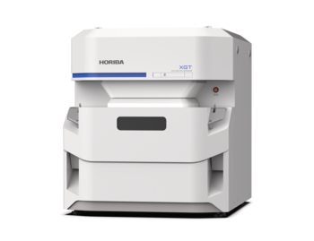
X-ray Analytical Microscope (Micro-XRF)
Do you have any questions or requests? Use this form to contact our specialists.