
Raman spectroscopy was named after Sir Chandrasekhara Venkata Raman (7 November 1888 – 21 November 1970), an Indian physicist born in the former Madras Province in India, who carried out ground-breaking work in the field of light scattering, which earned him the 1930 Nobel Prize for Physics.
During a voyage to Europe in 1921, Raman noticed the blue color of glaciers and the Mediterranean Sea. He was motivated to discover the reason for the blue color. Raman carried out experiments regarding the scattering of light by water and transparent blocks of ice which explained the phenomenon.
Raman employed monochromatic light from a mercury arc lamp which penetrated transparent material and was allowed to fall on a spectrograph to record its spectrum. He detected lines in the spectrum, which were later called Raman lines. He presented his theory at a meeting of scientists in Bangalore on 16 March 1928, and won the Nobel Prize in Physics in 1930. In Munich, some physicists were initially unable to reproduce Raman's results, leading to skepticism. However, Peter Pringsheim was the first German to reproduce Raman's results successfully and sent his spectra to Arnold Sommerfeld. Pringsheim was the first to coin the term "Raman effect" and "Raman lines."
Detection was either with a photographic plate or a photomultiplier tube. The photographic plate was sometimes mounted as close as a few millimeters from the focusing lens, producing an f/0.9 system! “Baked plates”, where a technique of heating the plates to increase sensitivity, was used. To detect a signal that is weaker than the excitation signal by at least 6 to 8 orders of magnitude is indeed a challenge.
Practically this means that the “wings” from the elastically scattered light (the laser wavelength) will overwhelm the desired signal at the shifted wavelengths. During the 30’s, 40’s and 50’s, the “problems” that were commonly experienced, fluorescence and stray light, were avoided because the samples were extensively purified (multiple distillations) after preparation; as much as 3 months for total preparation (particles in solution would produce a flash of light that would ruin the plate.) During the earliest period, spectra were typically recorded with prism spectrographs, Hg lamps, and photographic plates, integrating sometimes for days. Jobin & Yvon lenses were used already in triple prism-based model produced by Parisian company Huet.
When people began to study polycrystalline materials, problems of stray light and luminescence (from impurities) became overwhelming obstacles to the acquisition of high-quality spectra.
The first commercial Raman instrument using dispersive gratings and laser as an excitation source, was introduced as early as 1966, according to literature, following the advent of the laser in the 1960s.
First commercial Raman spectrometers at HORIBA
These first 2 systems, unveiled in 1967 were almost simultaneously delivered in 1968 by Coderg and Spex companies, incorporated later in the 80s in the Instruments SA group, now part of the Raman spectroscopy division of HORIBA.
The Spex system, called Spex 1401 Ramalog double monochromator, designed by Sergio Porto and Don Landon, was successfully used, as early as 1970 at GeorgiaTech Institute of Technology, to measure high quality spectra of lactalbumin protein crystals and compare them with lyophilized powders, and at Bell Laboratories to study solid state materials.
First installations of Coderg PH1 double monochromator (1968), and few years later Coderg T800 (1972) triple stage monochromator – designed by Delhaye under the guidance of Sergent-Rozey, Arie, Lescouarch, and Demol papers, are reported in the late 60s. In the literature, first publications using Raman Coderg spectrometers were published as early as 1968-69.
Holographic gratings: High quality spectra
Holographic Gratings - introduced commercially ca. 1972 - brought a drastic reduction of the stray light levels and eliminated ghosts. Thanks to the quasi perfection of those optical elements compared to their ruled grating cousins, Raman spectroscopy instrumentalists had a chance to improve the design of their spectrometers and the quality of the Raman spectra improved!
The Lirinord HG2S double monochromator was the first Raman spectroscopy instrument equipped with concave holographic gratings.
The 70s were, besides the disco-funk period in music, the time for the first developments of Raman microscopes, allowing probing minute quantities of samples rather than large volumes, and opening the door for imaging in Raman spectroscopy.
First Raman microscope leading to imaging: MOLE™
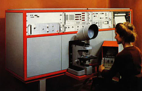
Professor P. Dhamelincourt and F. Wallart, from The LASIR laboratory in University of Lille, together with E. Da Silva, M. Leclercq, J. Barbillat, C. Allay and T. A. N’Guyen in France, and D. Landon in USA, all grouped under the Lirinord Instrument SA banner; worked out the first commercial Raman Laser microscope under the name MOLE, standing for Molecular Optical Laser Examiner, first described in 1966 by Delhaye and Migeon, who argued that a laser beam could be tightly focused at a sample, and the Raman light efficiently collected and transferred to a Raman spectroscopy system, without losses.
Calculations showed that increase in irradiance more than compensated for decrease in size of irradiated volume. The MOLE system was commercially released in 1976.
Advantages of the Microscope for Raman Sampling include:
Laser Focus: High numerical aperture enables tight focusing (~1 μm, diffraction limited)
Raman Collection: High numerical apertures enable collecting almost 2π steradians
Image Transfer: Correct optical coupling with spectrograph permits all Raman light to be transmitted by ~100 μm entrance slit and thus achieve high throughput
Fluorescence Rejection: Because the fluorescence can migrate away from the point of illumination, it can be rejected by the confocal optics.
Use of CCD detectors: High acquisition speed
The next big steps in Raman spectroscopy following the microscope introduction were the development of Charge Coupled Devices with improved sensitivity for Raman detection, and holographic Rayleigh filters.
The CCD cameras were applied as multichannel detectors providing spectral acquisition at least ten times faster than the then conventional instruments.
In order to take full advantage of the CCD technical breakthrough, several Raman companies including Spex, Jobin-Yvon and Dilor redesigned completely the spectrometers to achieve a flat field image over a larger width while correcting for optical aberrations.
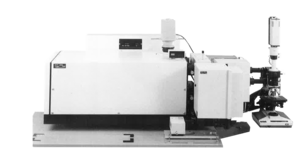
The optics in Czerny-Turner spectrographs were modified in order to achieve a focused image in a 1” flat field in the focal plane. The famous Jobin –Yvon U1000 in 1978, double additive Czerny-Turner monochromator, the Spex TripleMate™, commercialized in 1983, which made use of toroidal mirrors to correct astigmatism, whilst the MicroDil series (1981, 1983) used a camera lens to correct for coma and the Dilor XY (1986), used cylindrical lenses in front of the entrance slit.
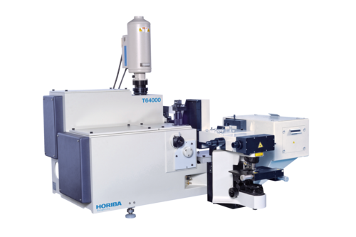
A few years later in 1988, Jobin Yvon T64000 triple stage Raman spectrometer, designed and commercially introduced by M. Leclercq, A. Thevenon and J. Oswalt, included the use of patented aberration correcting holographic (1990) gratings (PACH™) which, by the use of a hologram produced on an optical table with optics identical to those of the spectrograph, produced a holographic profile which corrects for astigmatism in Raman spectroscopy system.
Superhead InduRAM
First optical fiber coupled Raman probe for process applications designed with Professor Dao from Ecole Centrale Paris.
The SuperHead InduRAM is an optical fiber probe developed by HORIBA for Raman spectroscopy. Designed for industrial applications, it enables in situ and non-invasive chemical analysis, even in demanding environments. Its compact and robust design makes it suitable for various applications, including industrial reaction monitoring.
First compact single stage confocal Micro Raman: LabRAM™
The birth of the LabRAM Laboratory Raman spectroscopy microscope family followed soon after this, taking advantage of holographic high-quality Rayleigh filters, and high-sensitivity CCD array detectors.
At Pittcon conference in 1993, the LabRAM concept - first true confocal single stage Raman Microscope was launched.
The LabRAM has a focal length of 300 mm between the dispersing element, the grating, and the CCD detector, resulting in a spectral resolution of 2-4 cm-1 which is suitable for common applications with laser excitations between 400 and 800 nm.
The optical design received multiple improvements over the last 25 years, and innovations in terms of hardware and software under the engineering and guidance of E. Da Silva, M. Leclercq, B. Roussel, H-J. Reich, F. Adar, S. Morel, A. Whitley, E. Froigneux, Ph. De Bettignies, D. Tuschel, Yumei Pu and many others.
Below is the list of major steps in detection, filtering, imaging, computing, and hyphenation with other techniques that paved the way for state-of-the-art Raman spectroscopy systems today.
The various companies mentioned hereabove all joined HORIBA group in 1997, and their technologies were implemented over the years in the Raman spectroscopy family of products.
LabRAM HR
First deep UV 800 mm focal length spectrometer.
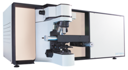
LabRAM HR (High Resolution) has an extended focal length of 800 mm leading to a spectral resolution which is around three times higher compared to the standard LabRAM. This increase in spectral resolution is also important for applications in the UV range or investigations such as stress measurement of semiconductor materials, polymorphism or similar where only small band shifts are investigated. Further flexibility is added to the LabRAM HR with the capability to incorporate a second detector (InGaAs) to extend the detectable region to NIR (up to 1700 nm). An important application for this is the combination of Raman with photoluminescence measurements, where you can compare Raman with absorption/emission processes based on electronic transitions.
LabRAM Infinity
First compact Raman microscope.
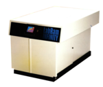
LabRAM IR2
First combination of FTIR Raman on single platform.
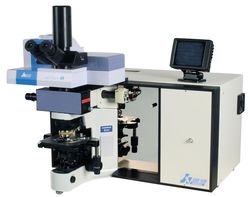
This new version is a combination of dispersive Raman and FTIR microscope, winner of the Gold Award for the best new product at PITTCON® 2002. The two complementary vibrational spectroscopic tools provide solutions to problems where the information from either technique is incomplete. SameSpot™ Technology allows both Raman and FTIR spectrum to be measured from the same place on the sample without having to move or transfer the sample.
ARAMIS
Fully automated Raman confocal microscope.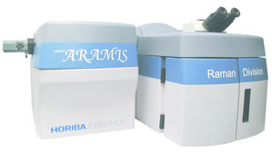
The LabRAM ARAMIS provides full system automation, including laser selection, grating exchange, and imaging functions, facilitating efficient and straightforward operation.
Open Space microscope
The advantage is open space and unlimited flexibility in sample adaptation whether you want to adapt Micro-cryostats, large DAC high pressure or high temperature cells, large samples (i.e. 300 mm wafers for inspection) or any other special sample arrangement!
Patented Line scan
Technology for fast mapping.
In Raman spectroscopy, line scanning involves illuminating and collecting data along a line across the sample rather than focusing on a single point or performing full area scans. This method enables faster data acquisition, as the system can capture detailed spectral information along the line in a single scan.
XploRA™
First ultra-compact and transportable bench-top Confocal Raman microscope below 40 kg.
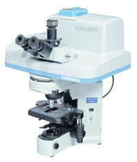
The XploRA turns a new page in microscopy. With an intuitive interface and full automation, Raman analysis has never been easier.
Chemical identification and chemical imaging can now be performed on solid or liquid samples at the touch of a button. Whether for routine sample identification, quantitative analysis or chemical imaging, the XploRA combines performance and simplicity in a cost-effective system. The light, compact design of the XploRA makes it easy to transport from lab to lab or for on-site analysis at archeological sites, crime scenes or in a mobile laboratory.
Patented SWIFT™
Fast Raman imaging.
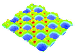
With point-by-point Raman mapping, a lot of the acquisition time is wasted in communication between the hardware and the software. Through continuous scanning of the sample, and new communication protocols between the scanning device and the CCD, SWIFT for the first time allows to reach acquisition times as low as 5ms/point, opening the door to quasi-instantaneous chemical imaging.
Patented Duoscan™
Innovation in Raman confocal scanning.
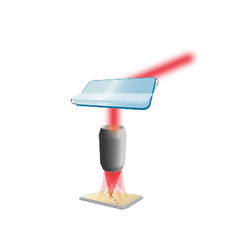
The DuoScan Imaging technology available on the instruments of the LabRAM Series introduces a new imaging mode, based on a combination of scanning mirrors that scan the laser beam across a pattern chosen by the operator: a line for linear profiles, or an area for two-dimensional mapping.
AccuRA
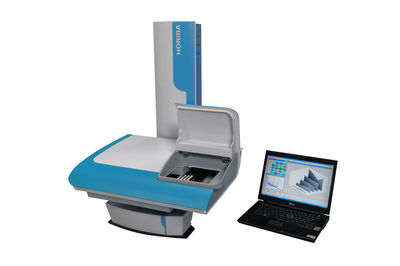
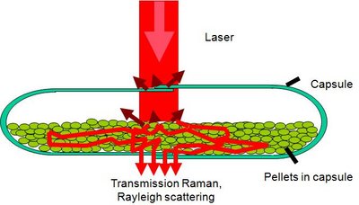
First stand-alone transmission Raman system.
Patented Ultra Low frequency filters on bench-top microscope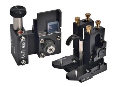
The ultra-low frequency (ULF) module allows Raman spectroscopic information in the sub-100 cm-1 region, with measurements below <10 cm-1 routinely available.
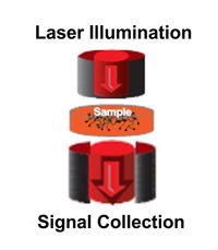
First combo Backscattered-Transmission Raman scattering
LabRAM HR Evolution with LabSpec6 software
Fully customer-oriented Raman Spectroscopy Software, simply powerful.

LabSpec 6 software offers great modularity with the exclusive LabStore Apps. Any user can configure their software according to their own needs. Efficiency and performance are combined with ease-of-use. The modern and intuitive design of the software makes it easier than ever to achieve a perfect Raman image.
First m-CARS
Spontaneous Raman imaging prototype delivered.
XploRA Nano
First “TERS-proven” NanoRaman imaging systems.
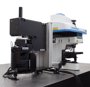
XploRA Nano is a versatile platform for physical and chemical characterization. Simultaneous AFM and spectroscopic measurements (Raman, Photoluminescence) are done thanks to high numerical aperture objectives from both top and side for best co-localized spatial resolution and best TERS collection efficiency.
Impact
Custom-made Ellipsometry and Raman under vacuum.
SWIFT XS™
First Ultrafast Raman imaging below 1 ms.
SWIFT XS integrates the HORIBA's newest EMCCD detector, combining unmatched speed and ultra-sensitivity. SWIFT XS is compatible with HORIBA's LabRAM HR Evolution and XploRA PLUS Raman microscopes.
First Stimulated / Spontaneous prototype for Raman imaging
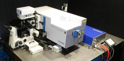
Particle Finder™
First Particle analysis embedded in Raman microscope.
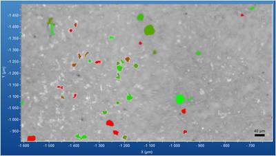
ParticleFinder, together with HORIBA Raman spectrometers, enables reliable and fast characterization of particles useful for pharmaceutical and chemical material development and quality control, forensic analysis, contaminant detection and geology.
MacroRAM™
First high-sensitivity cuvette-based benchtop Raman system.
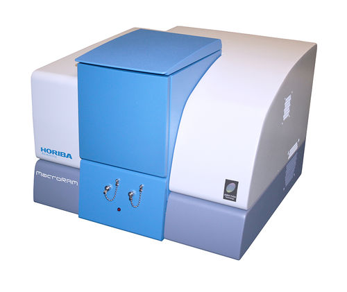
MacroRAM is an easy-to-use macro-Raman spectrometer for fast and reliable Raman analysis. Perfect for bulk analysis of solids, liquid solutions, powders, and gels, MacroRAM provides the flexibility and sensitivity to handle virtually any type of sample. With a compact and robust design including Class 1* laser safety, MacroRAM is ideal for use in most environments, from undergraduate teaching labs to industrial QC and manufacturing.
UVI 74X
First broadband Raman / PL achromatic objective high vacuum compatible.
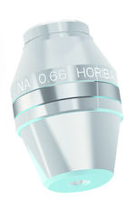
The objective UVI 74x is an achromatic broad range objective, based on Schwarzschild optical design. It is a fully reflective solution that removes all chromatic aberrations typically observed for UV objectives.
EasyNav™
First video image-based navigation experience combining fast autofocus and topography.
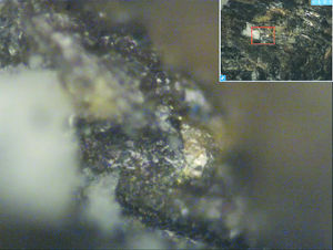
EasyNav is a package for LabSpec 6 allowing fast and easy navigation in-focus and in real-time, to identify the region of interest and obtain sharp, clear Raman chemical images. HORIBA NavMap™ + NavSharp™ + ViewSharp™ apps can be used together or separately to deliver a powerful user experience for all Raman users.
Patented SpecTop™ mode
Spec-Top is an original TERS imaging mode where TERS measurements are performed when the tip is in direct contact with the sample’s surface; transition between the pixels of the TERS map is performed in semicontact mode, which preserves the sharpness and enhancing properties of the AFM-TERS tip.
Integration of AIST-NT technology
HORIBA, global leader in Raman spectroscopy, announced in July 2017 the acquisition of AIST-NT technology, provider of innovative integrated scanning systems for nanotechnology. The combination of HORIBA’s Raman spectrometers with AIST-NT SPM technology allows HORIBA to offer academic research and industrial laboratories a range of integrated AFM-Raman systems with proven Tip Enhanced Raman Spectroscopy (TERS) solutions for the identification of chemicals and materials at the nanoscale. For the first time, an instrumentation company can provide a complete AFM-Raman solution; these range from the detector to the gratings, from the spectrometer to the AFM, fully manufactured by HORIBA.
SRGOLD technology
Thanks to Stimulated Raman Gain Opposite Loss Detection (SRGOLD), we achieved an increase of limit of detection of SRS imaging, making possible to achieve molecular imaging of biological samples. This analysis allows the spatial localization of chemical species of interest such as CH2 (lipid) and CH3 (protein) bonds to distinguish tissues where cell division is increased, characteristic of cancerous tissues.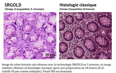
LabRAM Soleil™
A revolution in Raman imaging.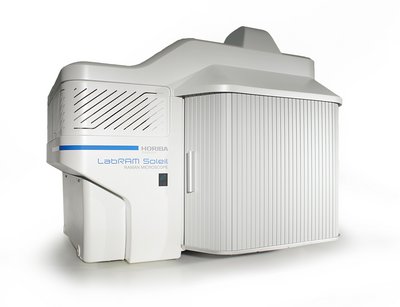
The LabRAM Soleil Confocal Raman Microscope is a cutting-edge instrument with advanced automation and imaging features. It offers precise molecular and structural characterization, with ultrafast imaging. Its compact design and laser-safe operation make it suitable for diverse applications, from material science to pharmaceuticals and nanotechnology. LabRAM Soleil facilitates comprehensive sample characterization and analysis with efficiency and precision, making it an indispensable tool for researchers and industrial users alike.
SmartSampling™
A new Smart way to do imaging.
SmartSampling offers incredible acquisition performance for high-resolution images, even for weak scatterers, in a fraction of the time required for standard maps. The technology uses an adaptative mapping step size approach to highlight the smallest microscopic details on the sample’s surface.
QScan™
Mapping any sample without constraints.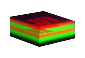
Unique to the LabRAM Soleil Raman multimodal microscope, this feature is compatible with all laser excitation wavelengths, from NUV to NIR and enables laser excitation and Raman / Photoluminescence collection from the same probed volume. QScan generates highly homogeneous laser lightsheet, enabling non-destructive confocal slicing of multilayer samples. It enables mapping without moving and true point-and-shoot operation, when one can directly acquire a spectrum by clicking anywhere in the video image.
χSTaiN™
The first integration of AI (Artificial Intelligence) in commercial Raman data processing solution.
χSTaiN is an innovative and intelligent tool for fully automated processing and analysis of Raman 2D images. It builds on HORIBA’s multidecade expertise in the analysis of spectral images through our global network of partners.
NanoGPS
Collaborative Correlative Microscopy Solution.
Patented NanoGPS technology provides a convenient way to create multiscale, multimodal correlative microscopy maps, with a registration accuracy that is limited only by the accuracy of the translation stage.
Acquisition of Process Instruments: from Lab to Process monitoring
HORIBA has strengthened its capabilities in industrial process monitoring by acquiring Process Instruments, Inc. (PI), a U.S.-based company known for its advanced technologies in process control and measurement. The acquisition aligns with HORIBA's strategy to expand its global presence in the industrial sector, enhancing its portfolio with PI's expertise in emission monitoring and process analytics. This move allows HORIBA to better meet the growing demand for reliable and precise monitoring technologies in various industries, including environmental and energy sectors.
ParticleFinder™ and IDFinder integration
A full solution for microplastics characterization.

ParticleFinder provides an integrated and customizable workflow for particle analysis. IDFinder enables the effortless creation and management of libraries, and identification of components from their Raman spectra in less than 100 milliseconds per spectrum.
LabRAM Odyssey Semiconductor
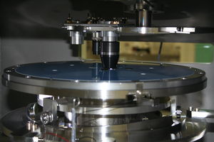
A dedicated system for full wafer characterization.![]()
The LabRAM Odyssey Semiconductor microscope is the ideal tool for photoluminescence and Raman imaging on wafers up to 300 mm diameter. The HORIBA best-seller true confocal microscope is equipped with automated 300 mm sample stage and objective turret to fit both needs of wafer uniformity assessment and defects inspection.
SignatureSPM
First Atomic Force Microscope (AFM) with built-in Raman/Photoluminescence spectrometer.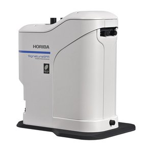
The SignatureSPM is an advanced multimodal characterization platform integrating an automated Atomic Force Microscope (AFM) with a Raman/Photoluminescence spectrometer. This integration enables true colocalized measurements of both physical and chemical properties of samples, providing comprehensive insights in a single, real-time analysis.
Tell us your anecdotes, your best paper, with your standard or custom-made Coderg, Spex, DILOR, Lirinord, Jobin-Yvon, Instrument SA, or HORIBA Raman spectroscopy system at info-sci.fr(at)horiba.com.
Masz pytania lub prośby? Skorzystaj z tego formularza, aby skontaktować się z naszymi specjalistami.