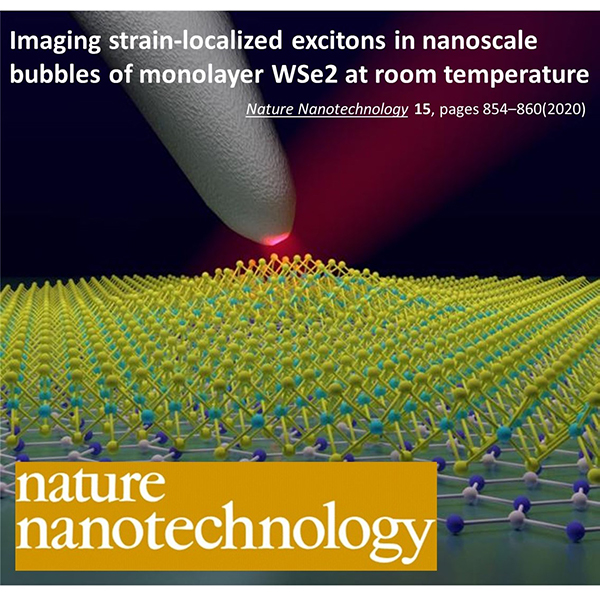
“Imaging strain-localized excitons in nanoscale bubbles of monolayer WSe2 at room temperature” Thomas P. Darlington, Christian Carmesin, Matthias Florian, Emanuil Yanev, Obafunso Ajayi, Jenny Ardelean, Daniel A. Rhodes, Augusto Ghiotto, Andrey Krayev, Kenji Watanabe, Takashi Taniguchi, Jeffrey W. Kysar, Abhay N. Pasupathy, James C. Hone, Frank Jahnke, Nicholas J. Borys & P. James Schuck Nature Nanotechnology 15, 854–86 (2020)
“Nanometer-Scale Uniform Conductance Switching in Molecular Memristors” Sreetosh Goswami, Debalina Deb, Agnès Tempez, Marc Chaigneau, Santi Prasad Rath, Manohar Lal, Ariando, R. Stanley Williams, Sreebrata Goswami, Thirumalai Venkatesan Advanced Materials 32, 42, 2004370 (2020)
“Uncovering topographically hidden features in 2D MoSe2 with correlated potential and optical nanoprobes” David Moore, Kiyoung Jo, Christine Nguyen, Jun Lou, Christopher Muratore, Deep Jariwala & Nicholas R. Glavin npj 2D Mater Appl 4, 44 (2020)
“Chemical Vapor Deposition of MoS2 for Energy Harvesting: Evolution of the Interfacial Oxide Layer” Tim Verhagen, Alvaro Rodriguez, Martin Vondráček, Jan Honolka, Sebastian Funke, Magda Zlámalová, Ladislav Kavan, Martin Kalbac, Jana Vejpravova, and Otakar Frank ACS Appl. Nano Mater, 3, 7, 6563–6573 (2020)
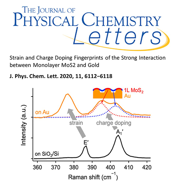
“Strain and Charge Doping Fingerprints of the Strong Interaction between Monolayer MoS2 and Gold” Matěj Velický, Alvaro Rodriguez, Milan Bouša, Andrey V. Krayev, Martin Vondráček, Jan Honolka, Mahdi Ahmadi, Gavin E. Donnelly, Fumin Huang, Héctor D. Abruña, Kostya S. Novoselov, and Otakar Frank J. Phys. Chem. Lett., 11, 15, 6112–6118 (2020)
“Strong Localization Effects in the Photoluminescence of Transition Metal Dichalcogenide Heterobilayers” Alvaro Rodriguez, Martin Kalbac, Otakar Frank arXiv:2010.06326 (2020)
“Radiative Decay of Dark Exciton Related Emission in a Sandwiched Monolayer WSe2 Revealed by Room Temperature Micro and Nano Photoluminescence” Mahfujur Rahaman, Oleksandr Selyshchev, Yang Pan, Ilya Milekhin, Apoorva Sharma, Georgeta Salvan, Sibylle Gemming, Dietrich R T Zahn arXiv:2006.04979 (2020)
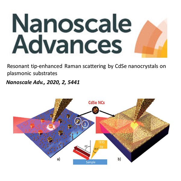
“Resonant tip-enhanced Raman scattering by CdSe nanocrystals on plasmonic substrates” Milekhin, M. Rahaman, K. V. Anikin, E. E. Rodyakina, T. A. Duda, B. M. Saidzhonov, R. B. Vasiliev, V. M. Dzhagan, A. G. Milekhin, A. V. Latyshev and D. R. T. Zahn Nanoscale Adv., 2020, 2, 5441-5449
“Picoscale control of quantum plasmonic photoluminescence enhancement at 2D lateral heterojunction” Zachary H Withers, Sharad Ambardar, Xiaoyi Lai, Jiru Liu, Alina Zhukova, Dmitri V Voronine arXiv:2001.10138 (2020)
“Observation of propagating surface plasmon polaritons by using functionalized tip‐enhanced Raman spectroscopy” Chawki Awada Chahinez Dab Jiawei Zhang Andreas Ruediger Journal of Raman Spectroscopy 51, 8, 1270-1277 (2020)
“Topography-induced variations of localized surface plasmon resonance in tip-enhanced Raman configuration” Azza Hadj Youssef, Jiawei Zhang, Andreas Dörfler, Gitanjali Kolhatkar, Alexandre Merlen, and Andreas Ruediger Optics Express 28, 9, 14161-14168 (2020)
“Sample induced intensity variations of localized surface plasmon resonance in tip-enhanced Raman spectroscopy” Jiawei Zhang, Azza Hadj Youssef, Andreas Dörfler, Gitanjali Kolhatkar, Alexandre Merlen, and Andreas Ruediger Optics Express 28, 18, 25998-26006 (2020)
“Tip-Enhanced Raman Nanospectroscopy of Smooth Spherical Gold Nanoparticles” Ashish Bhattarai, Zhihua Cheng, Alan G. Joly, Irina V. Novikova, James E. Evans, Zachary D. Schultz, Matthew R. Jones, and Patrick Z. El-Khoury J. Phys. Chem. Lett. 11, 5, 1795–1801 (2020)
“Suppressing Molecular Charging, Nanochemistry, and Optical Rectification in the Tip-Enhanced Raman Geometry” Chih-Feng Wang, Brian T. O’Callahan, Dmitry Kurouski, Andrey Krayev, Zachary D. Schultz, and Patrick Z. El-Khoury J. Phys. Chem. Lett. 2020, 11, 15, 5890–5895
“Direct Experimental Evidence of Hot Carrier-Driven Chemical Processes in Tip-Enhanced Raman Spectroscopy (TERS)” Rui Wang, Jingbai Li, Joel Rigor, Nicolas Large, Patrick Z. El-Khoury, Andrey Yu. Rogachev, and Dmitry Kurouski J. Phys. Chem. C 124, 3, 2238–2244 (2020)
“A Closer Look at Corrugated Au Tips” Ashish Bhattarai, Kevin T. Crampton, Alan G. Joly, Chih-Feng Wang, Zachary D. Schultz, and Patrick Z. El-Khoury J. Phys. Chem. Lett 11, 5, 1915–1920 (2020)
“In Situ Probing the Localized Optoelectronic Properties of Defective Monolayer WS2” Yi Yao, Fei Chen, Li Fu, Su Ding, Shichao Zhao, Qi Zhang, Weitao Su*, Xin Ding, and Kaixin Song J. Phys. Chem. C 124, 13, 7591–7596 (2020)
“Tip-Enhanced Multipolar Raman Scattering” Chih-Feng Wang, Zhihua Cheng, Brian T. O’Callahan, Kevin T. Crampton, Matthew R. Jones, and Patrick Z. El-Khoury J. Phys. Chem. Lett. 11, 7, 2464–2469 (2020)
“Spatio-Spectral Characterization of Multipolar Plasmonic Modes of Au Nanorods via Tip-Enhanced Raman Scattering” Ashish Bhattarai, Brian T. O’Callahan, Chih-Feng Wang, ShanYi Wang, and Patrick Z. El-Khoury J. Phys. Chem. Lett. 11, 8, 2870–2874 (2020)
“Comparable Enhancement of TERS Signals from WSe2 on Chromium and Gold” Andrey Krayev, Sergiy Krylyuk, Robert Ilic, Angela R. Hight Walker, Ashish Bhattarai, Alan G. Joly, Matěj Velický, Albert V. Davydov, and Patrick Z. El-Khoury J. Phys. Chem. C 124, 16, 8971–8977 (2020)
“Nanoscale Structural Characterization of Individual Viral Particles Using Atomic Force Microscopy Infrared Spectroscopy (AFM-IR) and Tip-Enhanced Raman Spectroscopy (TERS)” Tianyi Dou, Zhandong Li, Junjie Zhang, Alex Evilevitch, and Dmitry Kurouski Anal. Chem. 92, 16, 11297–11304 (2020)
“Gap-Mode Tip-Enhanced Raman Scattering on Au Nanoplates of Varied Thickness” Rui Wang, Zhe He, Alexei V. Sokolov, and Dmitry Kurouski J. Phys. Chem. Lett. 11, 10, 3815–3820 (2020)
“The Prevalence of Anions at Plasmonic Nanojunctions: A Closer Look at p-Nitrothiophenol” Chih-Feng Wang, Brian T. O’Callahan, Dmitry Kurouski, Andrey Krayev, and Patrick Z. El-Khoury J. Phys. Chem. Lett. 11, 10, 3809–3814 (2020)
“Elucidation of Photocatalytic Properties of Gold–Platinum Bimetallic Nanoplates Using Tip-Enhanced Raman Spectroscopy” Zhandong Li and Dmitry Kurouski J. Phys. Chem. C 124, 23, 12850–12854 (2020)
“Power-Dependent Dual Analyte Tip-Enhanced Raman Spectral Imaging” Brian T. O’Callahan, Ashish Bhattarai, Zachary D. Schultz, and Patrick Z. El-Khoury J. Phys. Chem. C, 124, 28, 15454–15459 (2020)
“Nanoscale Photocatalytic Activity of Gold and Gold–Palladium Nanostructures Revealed by Tip-Enhanced Raman Spectroscopy” Zhandong Li, Rui Wang, and Dmitry Kurouski J. Phys. Chem. Lett. 11, 14, 5531–5537 (2020)
“Spatially Resolved Mapping of Three-Dimensional Molecular Orientations with ∼2 nm Spatial Resolution through Tip-Enhanced Raman Scattering” Patrick Z. El-Khoury* and Edoardo Aprà J. Phys. Chem. C 124, 31, 17211–17217 (2020)
“Tip-enhanced Raman imaging of photocatalytic reactions on thermally-reshaped gold and gold–palladium microplates” Zhandong Li, Patrick Z. El-Khoury and Dmitry Kurouski Chem. Commun., 2021,57, 891-894
“Distilling Nanoscale Heterogeneity of Amorphous Silicon using Tip-enhanced Raman Spectroscopy (TERS) via Multiresolution Manifold Learning”Yang G, Li X, Cheng Y, Wang M, Ma D, Sokolov A, Kalinin S, Veith G, Nanda J Research Square; 2020. DOI: 10.21203/rs.3.rs-38466/v1.
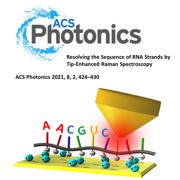
“Resolving the Sequence of RNA Strands by Tip-Enhanced Raman Spectroscopy” Zhe He, Weiwei Qiu, Megan E. Kizer, Jizhou Wang, Wencong Chen, Alexei V. Sokolov, Xing Wang, Jonathan Hu, and Marlan O. Scully ACS Photonics, (2020)
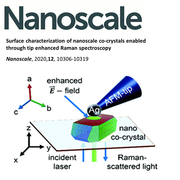
“Surface characterization of nanoscale co-crystals enabled through tip enhanced Raman spectroscopy” Jakob Hübner, Tanja Deckert-Gaudig, Julien Glorian, Volker Deckert and Denis Spitzer Nanoscale, 2020,12, 10306-10319 (2020)
“Formation Mechanism of Anisotropic RDX/TNT Core/Shell Nanoparticles and their Influence onto Nanodiamond Detonation Syntheses” Jakob Hübner, Vincent Pichot, Emeline Lobry, Tanja Deckert-Gaudig, Volker Deckert, Denis Spitze researchgate.net (2020)
“Facile and quantitative estimation of strain in nanobubbles with arbitrary symmetry in 2D semiconductors verified using hyperspectral nano-optical imaging” Thomas P. Darlington, Andrey Krayev, Vishal Venkatesh, Ravindra Saxena, Jeffrey W. Kysar, Nicholas J. Borys, Deep Jariwala, and P. James Schuck J. Chem. Phys. 153, 024702 (2020)
“Determining the Level and Location of Functional Groups on Few-Layer Graphene and Their Effect on the Mechanical Properties of Nanocomposites” Elizabeth J. Legge, Keith R. Paton, Magdalena Wywijas, Greg McMahon, Rory Pemberton, Naresh Kumar, Arun Prakash Aranga Raju, Craig P. Dawson, Andrew J. Strudwick, James W. Bradley, Vlad Stolojan, S. Ravi P. Silva, Stephen A. Hodge, Barry Brennan, and Andrew J. Pollard ACS Appl. Mater. Interfaces 12, 11, 13481–13493 (2020)
“Nanoscale characterization of plasma functionalized graphitic flakes using tip-enhanced Raman spectroscopy” Naresh Kumar, Sofia Marchesini, Thomas Howe, Lee Edwards, Barry Brennan and Andrew J. Pollard J. Chem. Phys. 153, 184708 (2020)
“Multi-technique physico-chemical characterization of particles generated by a gasoline engine: Towards measuring tailpipe emissions below 23 nm” C. Focsa, D. Duca, J. A. Noble, M. Vojkovic, Y. Carpentier, C. Pirim, C. Betrancourt, P.Desgroux, T. Tritscher, J. Spielvogel, M. Rahman, A. Boies, K. F. Lee; A. N. Bhave; S. Legendre, O. Lancry, P. Kreutziger, M. Rieker Atmospheric Environment 235, 117642 (2020)
“Deciphering tip-enhanced Raman imaging of carbon nanotubes with deep learning neural networks” Usant Kajendirarajah, María Olivia Avilés and François Lagugné-Labarthet Phys. Chem. Chem. Phys. 22, 17857-17866 (2020)
“Fabrication of a Biocompatible Mica/Gold Surface for Tip‐Enhanced Raman Spectroscopy” Dr. Xiao You, Clayton B. Casper, Emily E. Lentz, Prof. Dorothy A. Erie, Prof. Joanna M. Atkin ChemPhysChem 21, 188 – 193 (2020)
“Laser spectroscopic technique for direct identification of a single virus I: FASTER CARS” Volker Deckert, Tanja Deckert-Gaudig, Dana Cialla-May, Jürgen Popp,Roland Zell, Stefanie Deinhard-Emmer, Alexei V. Sokolov, Zhenhuan Yi, and Marlan O. Scully PNAS 117 (45), 27820-27824 (2020)