


PDF
2.58
MB

Helping you Release the Full Potential of your Extracellular Vesicles
Portfolio of Proven and Novel Characterization Techniques

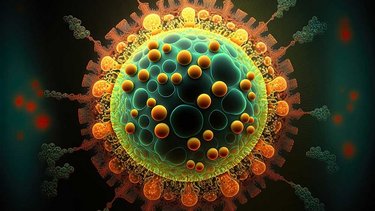
Extracellular vesicles (EVs) are small membranous structures that are released from a variety of cell types. These can be found in various bodily fluids, including blood, urine, and saliva. These are known to play critical roles in intercellular communication and can contain a variety of bioactive molecules, including proteins, lipids, and nucleic acids. Given their importance in both normal physiological processes and disease states, interest has increased in the analysis of EVs.
The first experiments in which EVs were studied began in the 80’s. The interest for these vesicles has been growing ever since, with an exponential development of the field over the past two decades.
EV analysis can involve a wide range of techniques that are essential for characterizing the size, concentration, and cargo of EVs, as well as for identifying the specific cell types that release it. Additionally, EV analysis can provide insights into the mechanisms by which EVs are taken up by recipient cells and the biological effects these induce.
Extracellular vesicles (EVs) can be broadly classified into three categories based on their biogenesis and size.
Exosomes: These are small EVs (30-150 nm) that are derived from the endosomal pathway. They are formed by the inward budding of the endosomal membrane and the subsequent fusion of multivesicular bodies (MVBs) with the plasma membrane. Exosomes carry various biomolecules, including proteins, lipids, and nucleic acids, and are involved in intercellular communication and cargo delivery.
Microvesicles: These are larger EVs (100-1000 nm) that are formed by the outward budding of the plasma membrane. Microvesicles carry a variety of bioactive molecules, including growth factors, cytokines, and signaling lipids. They are involved in intercellular communication, immune regulation, and blood clotting.
Apoptotic bodies: These are large EVs (1-5 µm) that are released by cells undergoing programmed cell death (apoptosis). Apoptotic bodies carry a diverse range of molecules, including chromatin, enzymes, and cellular organelles. These are involved in the clearance of apoptotic cells by phagocytes and the maintenance of tissue homeostasis.
We can consider small EVs (<200 nm) and medium/large EVs (<200 nm).
They are involved in various physiological and pathological processes, and researchers have extensively studied its potential applications in medicine and biotechnology. Extracellular vesicles have a wide range of uses in various, and their therapeutic potential is currently being explored in many preclinical and clinical studies.
Intercellular communication: EVs play an important role in intercellular communication by transferring biological molecules, such as proteins, lipids, and nucleic acids, between cells. This process can influence various physiological and pathological processes, including immune response, tissue regeneration, and cancer progression.
Diagnostic markers: EVs can serve as diagnostic markers for various diseases, as it contains biomolecules that reflect the physiological state of the parent cell. For example, circulating EVs have been shown to contain biomarkers for cancer and neurodegenerative diseases, and their analysis can aid in disease diagnosis and prognosis.
Drug delivery: EVs can be engineered to deliver therapeutic molecules, such as drugs or nucleic acids, to target cells. These offer several advantages over traditional drug delivery systems, including high biocompatibility, low immunogenicity, and the ability to cross biological barriers.
Tissue engineering: EVs can promote tissue regeneration and repair by modulating cellular behavior and signaling pathways. These have been used in various tissue engineering applications, including bone regeneration, cartilage repair, and wound healing.
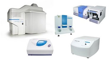
HORIBA Scientific instrumentation can help in the characterization of extracellular vesicles (EVs) in several ways:
HORIBA's nanoparticle tracking analysis (NTA) systems can accurately measure the size and concentration of EVs. NTA is a powerful tool for EV analysis, as it allows for the simultaneous measurement of particle size and concentration in real-time. Additionally, the use of fluorescent labeling with NTA enables the discrimination of different EV subpopulations based on its biomolecular content.
The zeta potential of EVs can provide valuable information about their surface charge and stability. HORIBA's zeta potential analyzers can measure the zeta potential of EVs, allowing researchers to understand how EVs interact with other biological structures. nanoPartica SZ-100V2 can also provide information about the size of extracellular vesicles.
Raman spectroscopy is a powerful analytical technique that can be used to identify the chemical composition of EVs. This technique is ideal for EV characterization, as it can provide high-resolution spectra with minimal sample preparation.
Raman spectroscopy can be combined with the AFM technique (TERS) for a more efficient analysis and to get nanoscale chemical and structural information and topography distribution, making the AFM-Raman platform a powerful tool.
A-TEEM fluorescence method quickly fingerprints molecules, differentiates exosome types, detects contaminants, and studies the biodistribution of extracellular vesicles. It is a non-destructive and label-free tool, insensitive to many common excipients while being hypersensitive to proteins and consequently to EVs. A-TEEM profiles provide better insights into protein signatures in complex matrices. This makes A-TEEM a valuable tool for QA/QC and process analysis.
The main interest of Surface Plasmon Resonance imaging is its ability to provide information about the presence of specific surface markers. This information can be used to distinguish different subpopulations of EVs, which may have different biological functions.
Providing Solutions for the Life Science Field Using Spectroscopic Analyzers (Readout No. E55)
READOUT is a technical journal issued by HORIBA.
Do you have any questions or requests? Use this form to contact our specialists.
