
Dynamic Image Analysis (DIA) is an advanced measurement technique that continuously captures images of particles in motion. By extracting particle outlines from these acquired images, the system calculates precise particle size distributions and shape parameters such as circularity and aspect ratio. Thanks to its ability to measure a vast number of particles in a short timeframe, DIA is widely utilized for particle size evaluation and morphological quality control.
In the field of image analysis, there are two primary methods: Static Image Analysis, which involves capturing images of stationary particles (e.g., on a slide), and Dynamic Image Analysis, which continuously captures high-speed images of particles actively moving through a measurement zone.
Static imaging allows flexible adjustment of the time required for image acquisition, making it easy to capture detailed images and combine them with component analysis such as elemental analysis. However, capturing a large number of particles requires repeatedly changing the imaging area and acquiring images, which is time-consuming. Dynamic imaging allows for continuous image acquisition of moving particles, making it relatively easy to capture a large number of images and images of many particles.
Depending on the distribution shape, obtaining a particle size distribution with sufficient accuracy is said to require information from tens of thousands of particles. Therefore, dynamic imaging is generally more suitable for creating particle size distributions.
Since dynamic image analysis covers a wide size range, it is often used in applications that have historically relied on sieving. By selecting appropriate size parameters, the results can closely match sieve results. In addition, it enables quick and easy analysis and provides additional information on particle shape.
For details, see Steps from Observed Image to Particle Size Distribution.
Figure of an optical microscope observation system
Static Imaging: The particles being measured are stationary, while the observation area moves.
Example: Optical microscope
Figure of a dynamic imaging observation system
Dynamic Imaging: Particles to be measured move through a fixed observation area.
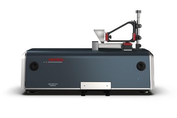
Example: Dynamic imaging particle size distribution analyzer
Dynamic image analysis uses many of the same steps as static image analysis. However, sample preparation is completely different because the sample is moving during measurement. Many of the image-processing steps used in static image analysis are also used in dynamic systems, but it is less common for the operator to actively select the functions being used.
Figure: Captured particle image and particle detection
Steps from Observed Image to Particle Size Distribution
Typically, a single image contains multiple particles, and particle regions can be separated from non-particle regions. Depending on the optical system, particles may appear bright (white) or dark (black). Particles are detected within an image by distinguishing them from the background based on the difference in their brightness.
Detected particles are represented as groups of pixels, as shown in the image here. Therefore, particle-size accuracy is determined by the pixel size (i.e., the actual length represented by one pixel). In contrast, the minimum detectable particle diameter depends on both the pixel size and the optical system. For example, if a particle image is captured with an optical system that has a 5 μm resolution and a pixel size of 100 nm, the particle size can be measured with an accuracy of approximately 100 nm, while the minimum detectable particle diameter is approximately 5 μm.
Particle size or shape parameters can be calculated from the image information obtained through particle detection. For example, to determine the Feret diameter (see below), the number of pixels in the longest horizontal portion is counted, and the actual length is calculated using the optical system's magnification. To determine the equivalent circle diameter, the particle area is first calculated from the number of pixels classified as the particle (using the optical system’s magnification), and then the diameter of a circle with the same area is calculated.
The particle size distribution determined by general image analysis methods is typically presented as a histogram, with particle size (or another per-particle parameter) on the horizontal axis and the number of particles on the vertical axis.
Size / Shape parameters
There are many methods for extracting features from images containing detected particles. Common parameters are listed below.
Equivalent circle diameter: From the area of the particle, calculate the particle diameter that can be replaced in a circle having the same area.
Aspect ratio: Calculate the ratio of the width and length of the minimum-area rotated rectangle circumscribing the particle.
When the values of the width and length are equal, as in a perfect circle, the result is 1. Depending on which axis is used as the denominator, the value can range from zero to 1, or from 1 to infinity. For ease of use, the length is often used as the denominator so that the value ranges from zero to 1.
Circularity: To ensure that the circularity is equal to 1 for a perfect circle, calculate it by multiplying the ratio of the area to the square of the perimeter by a constant.
Feret diameter (Feret max, also called caliper diameter): The maximum distance between two parallel lines that touch the particle outline.
Dynamic image analysis is a technique where particles are continuously passed in front of a camera and imaged in real time. This allows for rapid measurement of particle size distribution and particle shape, providing more comprehensive information compared to traditional sieve analysis.
Static image analysis involves imaging particles that are stationary, typically on a microscope slide. Dynamic image analysis, on the other hand, measures particles as they move through the observation area. Dynamic methods are ideal for analyzing large numbers of particles quickly and for real-time monitoring.
Dynamic image analysis is well-suited for powders, granules, and suspensions where large numbers of particles need to be measured, and statistical analysis of particle size distribution and shape is required. It is widely used in industries such as pharmaceuticals, food, ceramics, and mining.
Particle shape parameters (such as aspect ratio, circularity, and sphericity) significantly affect the physical properties and performance of materials. For example, even if powders have the same particle size, those with a higher degree of circularity have the characteristic of having higher fluidity as a powder. Image analysis enables accurate measurement of these parameters, supporting quality control and process optimization.
The larger limit is determined by the size that exceeds the angle of view. The smaller limit is determined by both the size of one pixel and the optical resolution. The size of one pixel also affects how accurately particles can be measured.
Image analysis provides not only particle size distribution, but also detailed shape information, offering a more complete characterization than sieve analysis. Furthermore, finer particle size fractions allow for higher resolution particle size distributions to be obtained. It is also better suited for fine particles and automated measurements.
The larger limit is determined by the size that exceeds the angle of view. The smaller limit is determined by both the size of one pixel and the optical resolution. The size of one pixel also affects how accurately particles can be measured.
For dynamic image analysis, samples are typically dispersed in a liquid or fed dry to ensure uniform distribution. Proper dispersion and dilution are essential for accurate measurement.
Common parameters include equivalent circle diameter, maximum Feret diameter, aspect ratio, and circularity. These are used for physical characterization and process control.
Image analysis measures individual particle size and shape directly, while laser diffraction estimates particle size distribution based on light scattering. Each method has advantages depending on the application. When comparing, it is important to pay attention to the particle size basis (whether it is based on number or volume).
HORIBA’s Partica uses multiple cameras to perform wide-range dynamic image analysis and simultaneously measures particle size distribution using laser diffraction. This allows you to obtain size information from two measurement technologies at once, as well as shape information from an image analysis.
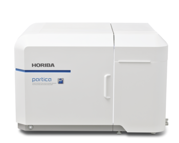
Laser Diffraction and Dynamic Imaging Particle Size and Shape Analyzer
Dynamic Image Analysis
Masz pytania lub prośby? Skorzystaj z tego formularza, aby skontaktować się z naszymi specjalistami.


