


PDF
11.05
MB

Fluorescent Bioprobes e-book
Our e-book provides you with an overview of the fluorescent bioprobes characterization. We explain and detail the analytical solutions, including fluorescence, particle analysis, Raman, and more.

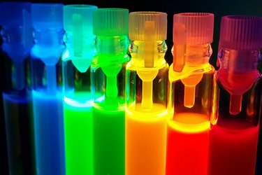
Fluorescent bioprobes are compounds that absorb light at a given wavelength and emit light at a higher wavelength, producing fluorescence in various colors. Their use in biological labeling and staining began as early as the 1930s, and the research community has come a long way since then. Biologists now have at their disposal a plethora of fluorescent bioprobes with different colors, structural properties, and functionalization...
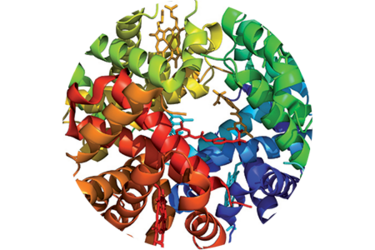
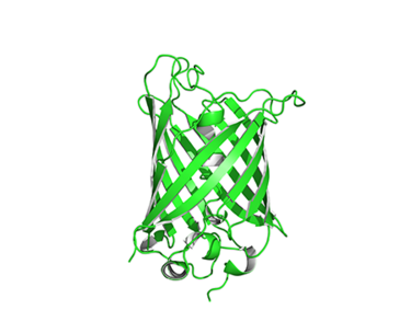
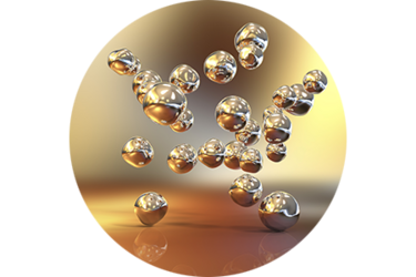
The large variety of fluorescent probes, such as organic dyes, biological biotags and fluorescent nanostructures, are intensively studied due to their inherent suitability for a wide range of life science applications.
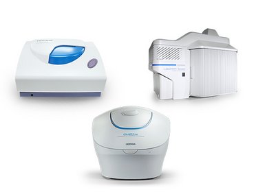
Thanks to the complementary nature of its analytical solutions, HORIBA can be your single partner to support you at various steps in your bioprobe synthesis and use.
This unique offer covers fluorescence, particle analysis, AFM, Raman and XRF characterization techniques, helping to gain valuable insight into optical properties, size, morphology and composition of your bioprobes.
Fluorescent probes are compounds that absorb light at a given wavelength and emit light at a higher wavelength, producing fluorescence in various colors. Their use in biological labeling and staining began as early as the 1930s, and the research community has gone a long way since then. Biologists now have at their disposal a plethora of fluorescent bioprobes with different colors, structural properties and functionalization...
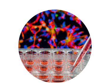
They are employed in a wide range of research applications. These include: Antibody labels to indicate the presence of a target antigen in a sample via immunofluorescence, reporter molecules when introducing a gene construct into a cell, stains for the quantitative measurement of nucleic acids, and indicators of cell health.
There is an increasing need for characterization tools during both the development and the use of the most common families of bioprobes, whether to determine their optical emission profiles, measure their size, or assess their elemental composition or molecular signature.
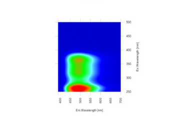
Fluorescence spectroscopy is the most commonly used technique to study fluorescent bioprobes to investigate their intrinsic properties and how they change depending on the physico-chemical environment.
For a full understanding of their properties, other techniques such as particle characterization and Raman spectroscopy are widely used for supplementary information. Our instruments are used by scientists all over the world to obtain rapid feedback on the best direction to follow to improve their processes.
Fluorescence enables evaluation of biotag properties throughout the synthesis process, leading to a better understanding of
their behavior and potential interactions with biological targets.
The optical properties of fluorescent nanoparticles can be fine-tuned by their size, which is a key parameter that determines the spectral position and purity of fluorescence.
Absorption, distribution, metabolism, excretion and toxicity depend on multiple factors such as size, charge and concentration.
Raman spectroscopy is an important tool for investigating the structural and compositional characteristics of biomolecules.
It can be used in life science for hyperspectral molecular imaging of cells and tissue and medical diagnostics. In the case of fluorescent probes, this technique can provide complementary information about composition and purity.
The atomic force microscope (AFM) is ideally suited for characterizing fluorescent nanostructures. It enables 3D visualization and provides information on many physical properties including size, morphology, surface texture and roughness. Statistical information, including size, surface area, and volume distributions, can be determined, as well.
Micro-XRF is a powerful, non-destructive analytical technique that can provide supplementary information for bioprobe characterization. The elemental mapping capabilities of this technique enable the detection of major elements, suitable to quickly detect and identify contaminants.
Read what scientists are saying about working with HORIBA instruments:
Ramsay, H., Simon, D., Steele, E., Hebert, A., Oleschuk, R. D., & Stamplecoskie, K. G. (2018). The power of fluorescence excitation–emission matrix (EEM) spectroscopy in the identification and characterization of complex mixtures of fluorescent silver clusters. RSC advances, 8(73), 42080-42086.
Using the power of A-TEEM spectroscopy, this study demonstrates that the A-TEEM fingerprint is able to resolve unique fluorescence properties in different silver clusters. The authors also used parallel factor analysis (PARAFC) to support the presence of three distinct compounds from an A-TEEM fingerprint that consisted of a complex mixture of silver clusters. This work also benefited from the extended NIR detection of the CCD inside Duetta, since some components emitted above 850 nm.
From this work, the authors concluded that “Based on these findings, EEM spectroscopy can be implemented as a powerful technique for determining the purity of complex mixtures, especially when other techniques, including mass spectrometry, fail to provide adequate characterization of a given material.” Dr Kevin Stamplecoskie, Assistant Professor Materials and Physical
This publication is a compelling testimonial to the power of A-TEEM spectroscopy as an analytical technique, and points to a valuable and significant benefit that Duetta provides with its unique A-TEEM molecular fingerprinting technology.
Altinolcek, N., Battal, A., Tavasli, M., Peveler, W. J., Holly, A. Y., & Skabara, P. J. (2020). Synthesis of novel multifunctional carbazole-based molecules and their thermal, electrochemical and optical properties. Beilstein journal of organic chemistry, 16(1), 1066-1074.
"Work in my group focusses on developing new chemical probes and sensors - in particular we synthesise luminescent small molecules and nanomaterials with bespoke targeting properties for tacking various biological challenges. One area of interest is creating sensing arrays where we exploit cross-reactivity amongst a set of luminescent sensors to ‘fingerprint’ a target protein, using this approach, and more traditional binding assays, we are developing diagnostic tests for liver fibrosis.
We use the HORIBA Duetta to evaluate our luminescent materials as we synthesise and modify them, and appreciate the really fast measurements enabled by the CCD detector. We undertake EEM measurements to study mixtures of luminescent sensors and hope to use the Peltier stage in future to examine reaction kinetics at different temperatures with both absorption and fluorescence simultaneously. Other instrument users carry out research on everything from photovoltaic and LED materials to water pollution, demonstrating how versatile the measurements can be!" Dr William Peveler, LKAS Research Fellow
This publication points out how the EEM measurements on the Duetta can streamline developing new chemical probes through fast measurements.
Get an overview of our customers' applications through stories from our Science in Action series showcasing HORIBA solutions for fluorescent bioprobes analysis:
HORIBA proposes a collection of application notes and webinars featuring our analytical solutions for fluorescent bioprobes characterization.
Quantum Dots
Organic Dyes
Carbon nanotubes
Metallic nanoclusters
Watch this presentation to learn about how the characterization of novel fluorophores can be made faster, easier, and more complete using the Duetta.

This talk gives an overview of HORIBA’s analytical solutions to help with the development of tailored and stable emitting bioprobes, in particular through the optimization of their chemical synthesis, with a focus on Fluorescence Spectroscopy and Particle Characterization.
Máte nějaké dotazy nebo požadavky? Pomocí tohoto formuláře kontaktujte naše specialisty.