XploRA Nano

AFM-Raman XploRA Nano

AFM-Raman pour l'imagerie physique et chimique

Système entièrement intégré basé sur le microscope à sonde locale de pointe SmartSPM et le micro-spectromètre Raman XploRA.

Compact, entièrement automatisé et facile à utiliser, le XploRA Nano concentre toute la puissance de la technologie AFM-Raman dans un système complet et abordable, mettant l'imagerie TERS à la portée de tous. Système TERS éprouvé.

Segment: Scientific
Fabricant: HORIBA France SAS

Plateforme d'analyse multi-échantillons

Il est possible d'effectuer des mesures à l'échelle macro, micro et nanométrique sur la même plateforme.

Facilité d'utilisation

Fonctionnement entièrement automatisé pour des mesures en quelques minutes.

De véritables performances confocales

Haute résolution spatiale, étapes de cartographie automatisées, options complètes de visualisation au microscope.

Collecte haute efficacité

Détection Raman par le dessus et par le côté pour optimiser la résolution et le rendement lors des mesures colocalisées et exaltées par effet de pointe (Raman et photoluminescence).

Haute résolution spectrale

Résolution spectrale ultra performante, plusieurs réseaux avec commutation automatique, analyse dans une large gamme spectrale pour Raman et PL.

Haute résolution spatiale

Résolution spectroscopique à l'échelle nanométrique (jusqu'à 10 nm) grâce aux spectroscopies optiques exaltées par effet de pointe (Raman et photoluminescence).

Multi-technique/Multi-environnement

Plusieurs modes SPM sont disponibles : modes AFM, conducteur et électrique (cAFM, KPFM), STM, cellule liquide et environnement électrochimique, cartographie chimique par TERS/TEPL, etc. Contrôle total sur les 2 instruments depuis un poste de travail avec un logiciel de contrôle puissant, possibilité d'utiliser le SPM et le spectromètre simultanément ou de façon indépendante

Robustesse/Stabilité

Scanners AFM à haute fréquence de résonance, extrêmement silencieux Haute performance sans isolation active contre les vibrations.

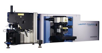
Scanner et base SmartSPM

Plage de balayage d'échantillon : 100 µm x 100 µm x 15 µm (± 10 %)

Type de balayage par échantillon : Non-linéarité XY 0,05 % ; non-linéarité Z 0,05 %

Bruit : 0,1 nm RMS dans la dimension XY sur une largeur de bande de 200 Hz avec les capteurs capacitatifs activés ; 0,02 nm RMS dans la dimension XY sur une largeur de bande de 100 Hz avec les capteurs capacitatifs désactivés ; < 0,04 nm RMS dans la dimension Z sur une largeur de bande de 1 000 Hz avec le capteur capacitatif

Fréquence de résonance : XY : 7 kHz (sans charge) ; Z : 15 kHz (sans charge)

Mouvement X, Y, Z : contrôle numérique en boucle fermée pour les axes X, Y, Z et plage d'approche Z motorisée 18 mm

Taille d'échantillon : 40 x 50 mm max., épaisseur 15 mm

Positionnement des échantillons : plage de positionnement motorisé des échantillons 5 x 5 mm

Résolution de positionnement : 1 µm

Tête AFM

Longueur d'onde du laser : 1 300 nm, sans interférence avec le détecteur spectroscopique

Alignement : alignement entièrement automatisé du levier et de la photodiode

Accès à la sonde : libre accès à la sonde pour des manipulateurs et des sondes externes supplémentaires
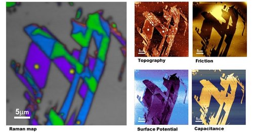
Modes de mesure SPM

AFM contact dans l'air (en milieu liquide en option) ; AFM contact intermittent dans l'air (en milieu liquide en option) ; AFM non-contact ; imagerie de phase ; microscopie à force latérale (LFM) ; modulation de force ; AFM conductrice (en option) ; microscopie à force magnétique (MFM) ; sonde de Kelvin (microscopie à potentiel de surface, SKM, KPFM) ; microscopie capacitive et à force électrique (EFM) ; mesures de courbe de force ; microscopie à force piézoélectrique (PFM) ; nanolithographie ; nanomanipulation ; STM (en option) ; cartographie du photocourant (en option) ; mesures de la caractéristique volt-ampère (en option)

Modes de spectroscopie

Imagerie et spectroscopie confocales Raman, de fluorescence et de photoluminescence

Spectroscopie Raman exaltée par effet de pointe (TERS) en modes AFM, STM et force de cisaillement

Photoluminescence exaltée par effet de pointe (TEPL)

Microscopie et spectroscopie optiques en champ proche (NSOM/SNOM)

Unité AFM conductrice (en option)

Plage de courant :  100 fA ÷ 10 µA ; 3 plages de courant (1 nA, 100 nA et 10 µA) commutables par logiciel

Accès optique

Possibilité d'utiliser simultanément un objectif planapochromatique supérieur et latéral : jusqu'à 100x, NA = 0,7 par le dessus ou le côté ; jusqu'à 20x et 100x simultanément

Scanner d'objectif piézoélectrique en boucle fermée pour un alignement laser spectroscopique ultrastable à long terme : plage 20 µm × 20 µm × 15 µm ; résolution : 1 nm

Spectromètre

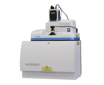
Micro-spectromètres compacts XploRA Plus entièrement automatisés, fonctionnant comme un microscope micro-Raman autonome

Gamme de longueurs d'onde : de 60 cm-1 à 4 000 cm-1

Réseaux : 4 réseaux sur une tourelle contrôlée par ordinateur (600, 1 200, 1 800 et 2 400 g/mm)

Automatisation : fonctionnement entièrement motorisé, contrôlé par logiciel

Détection

Gamme complète de détecteurs CCD et d'EMCCD.

Sources laser

Longueur d'onde standard : 532 nm, 638 nm, 785 nm.

Automatisation : fonctionnement entièrement motorisé, contrôlé par logiciel

Logiciels

Pack logiciel intégré avec SPM complet, spectromètre et outil de contrôle d'acquisition de données, comprenant une suite d'analyse et de traitement des données spectroscopiques et SPM (adaptation, déconvolution et filtrage de spectre inclus). Des modules en option proposent une suite d'analyse univariée et multivariée (PCA, MCR, HCA, DCA) et des fonctionnalités de détection de particules et de recherche spectrale.

TERS Characterization of Lipid Nanotubes as Carbonaceous Material for Electrodes
For thirty years there has been a research focused onto carbonization of 3D structures especially to be employed in electronic applications. These structures are prepared with the help of lithography and pyrolyzed afterwards. For making features below 100 nm, a bottom-up approach using lipids nanotubes is attempted.
TERS Characterization of Single- to Few-Layer Ti₃C₂Tₓ MXene
MXenes is the largest and fastest growing 2D materials. They have unique properties such as good conductivity and a hydrophilic surface. The control of nanoscale composition would ultimately allow for engineering properties locally, gaining more control over the 2D material-based systems.
TERS on Functionalized Gold Nanostructures for Nano-scale Biosensing
Surface-enhanced Raman scattering (SERS) is a powerful plasmonics-based analytical technique for biosensing. SERS effect relies on nanostructures that need to be designed to maximize enhancement factors and molecular specificity. In addition to numerical modeling, an analytical tool capable of imaging localized enhancement would be an added value.
TERS Characterization of phospholipid bilayers and detection of nanoparticles
Phospholipid bilayers, major constituents of membranes act as a barrier of selective permeability for the nanoparticles now largely into our environment. Studying the interactions between nanoparticles and cellular membranes requires a molecular chemical probe with nanometer resolution capability.
TERS Characterization of Graphene Nanoribbons
Graphene is foreseen for a handful of electronic and optoelectronic nano-devices. Making nano-devices out of graphene requires nanopatterning. Determining the quality of patterned graphene is essential and the detection of defects demands a sensitive chemical nano-characterization tool.
TERS Characterization of Explosive Nanoparticles
It is not yet understood how co-crystal nanoparticles (co-crystallinity combined with nanostructuring) have superior properties to single compound crystals. Only a technique capable of probing single nanoparticles can bring answers.
c-AFM and in operando TERS & µRaman Characterization of Molecular Switching in Organic Memristors
Emergence of organic memristors has been hindered by poor reproducibility, endurance stability scalability and low switching speed. Knowing the primary driving mechanism at the molecular scale will be the key to improve the robustness and reliability of such organic based devices.
Correlated TERS and KPFM of Graphene Oxide Flakes
Visualizing the distribution of structural defects and functional groups present on the surface of two-dimensional (2D) materials such as graphene oxide challenges the sensitivity and spatial resolution of most advanced analytical techniques.
AFM-TERS measurements in a liquid environment with side illumination/collection
Atomic Force Microscopy (AFM) associated to Raman spectroscopy has proven to be a powerful technique for probing chemical properties at the nanoscale. TERS in liquids will bring promising results in in-situ investigation of biological samples, catalysis and electrochemical reactions.
Characterization of Nanoparticles from Combustion Engine Emission using AFM-TERS
A new concern for human health is now raised by sub-23 nm particles emitted by on-road motor vehicles. Beyond measuring particle number and mass, it is also critical to determine the surface chemical composition of the nanoparticles to understand the potential reactivity with the environment.
Correlated TERS, TEPL and SPM Measurements of 2D Materials
Many challenges remain before the promise of 2D materials is realized in the form of practical nano-devices. An information-rich, nanoscale characterization technique is required to qualify these materials and assist in the deployment of 2D material-based applications.
Characterization of Carbon Nanotubes Using Tip-Enhanced Raman Spectroscopy (TERS)
The use of TERS to reveal the defects density in the structure of CNTs is of interest for a better understanding of the electrical properties of the devices made with such nano-objects. Not only defects concentration but also local chirality changes from the different radial breathing modes, pressure effect and strain distribution can be studied at the single carbon nanotube level through TERS.
Characterization of MoS2 Flakes using TEOS
Both TEPL and TERS images are well correlated with AFM morphological images obtained simultaneously, and all are consistent in revealing the nature (number of layers) of MoS2 flakes. Upon deconvolution, the TEPL signal is even capable of revealing local inhomogeneities within a MoS2 flake of 100 nm size. Kelvin probe measurement supports TEPL and TERS measurements and adds to the power of such tip-enhanced combinative tools. TEOS characterization of 2D materials is likely to contribute to further deployment of these materials into commercial products through a better understanding of their electrical and chemical properties at the nanoscale.

Request for Information

Do you have any questions or requests? Use this form to contact our specialists.

* These fields are mandatory.

Related products

LabRAM Odyssey
LabRAM Odyssey

Imagerie Confocale Raman et Spectromètre à Haute Résolution

LabRAM Odyssey Nano
LabRAM Odyssey Nano

AFM-Raman pour l'imagerie physique et chimique

LabRAM Soleil
LabRAM Soleil

Microscope Confocal Multimodal Raman

LabRAM Soleil Nano
LabRAM Soleil Nano

Nanoscopie corrélative directe en temps réel

OmegaScope
OmegaScope

Plateforme optique AFM

TRIOS
TRIOS

Couplage optique AFM polyvalent

XploRA Nano
XploRA Nano

AFM-Raman pour l'imagerie physique et chimique

XploRA™ PLUS
XploRA™ PLUS

Spectromètre Raman – Microscope confocal Raman

LabRAM Odyssey Nano
LabRAM Odyssey Nano

AFM-Raman pour l'imagerie physique et chimique

LabRAM Soleil Nano
LabRAM Soleil Nano

Nanoscopie corrélative directe en temps réel

ProteusQ
ProteusQ

Scanning NV Magnetometry

XploRA Nano
XploRA Nano

AFM-Raman pour l'imagerie physique et chimique

Corporate