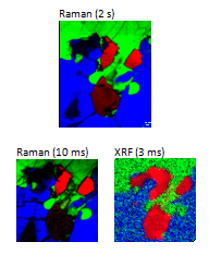

A polished granite section was analysed using both Raman and micro-XRF. The top Raman image was acquired with 2 s per point, and shows the distribution of FeS (red), SiO2 (green) and (K,Na)AlSi3O8 (blue).
On the bottom left, an ultra-fast SWIFT™ image is shown, acquired with 10 ms over the central portion of the original image. The same area was imaged (right) with the XGT-7000 micro-XRF analyzer, with a 10 µm X-ray beam and 3 ms per point acquisition. The XRF image shows the presence of elements Fe and S (red), Si (green) and K (blue).
Dr George J. Havrilla (Los Alamos National Laboratory, USA) is kindly thanked for providing this data. Data processing was carried out with the kind assistance from Jeremy Shaver (Eigenvector Research Inc., USA)