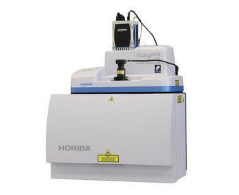
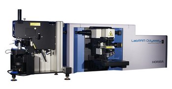
A família definitiva de microscópios Raman
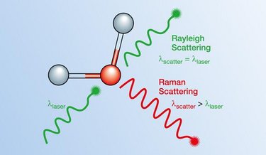
HORIBA é líder mundial em espectroscopia Raman, beneficiando-se de mais de 50 anos de inovação na técnica. A microscopia Raman é uma técnica que permite a análise química rápida e não destrutiva de sólidos, pós, líquidos e gases – hoje, a espectroscopia Raman é utilizada em diversas áreas, desde a pesquisa fundamental até soluções aplicadas.
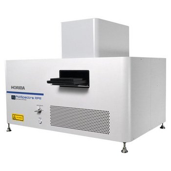
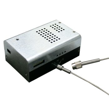
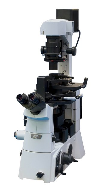
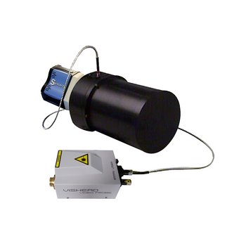
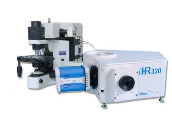
Oferecemos soluções completas de espectroscopia Raman para medições analíticas, pesquisa Raman, Raman UV, controle de qualidade/garantia de qualidade e aplicações industriais. Isso inclui espectrômetros Raman, sistemas Raman híbridos (como AFM-Raman), sistemas Raman modulares, analisadores Raman de transmissão, espectrômetros Raman dedicados para processos in situ e instrumentos Raman miniaturizados para fabricantes OEM de alto volume.
Os entusiastas da espectroscopia Raman podem obter novas informações sobre aplicações, produtos, webinars, artigos, etc., assinando a newsletter Raman XPerience e consultando edições anteriores.