Particle Analysis in Vaccine Manufacturing and Development

Size matters in vaccine delivery systems. Nanoparticles smaller than 200 nm generally present a greater immunogenic response than micro-particles larger than 1 micron.
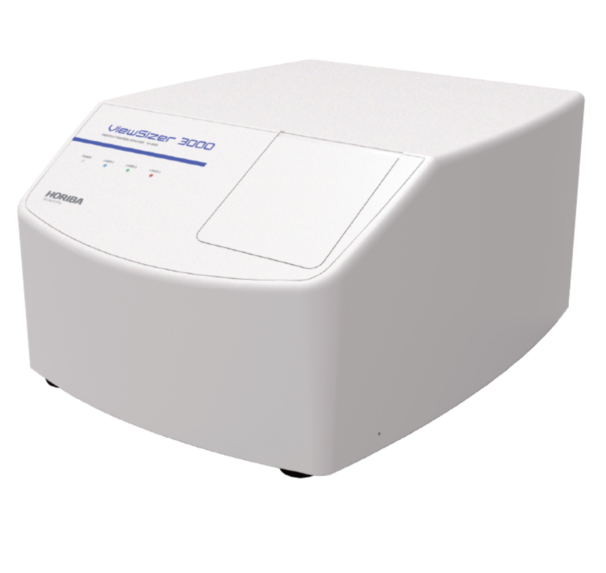
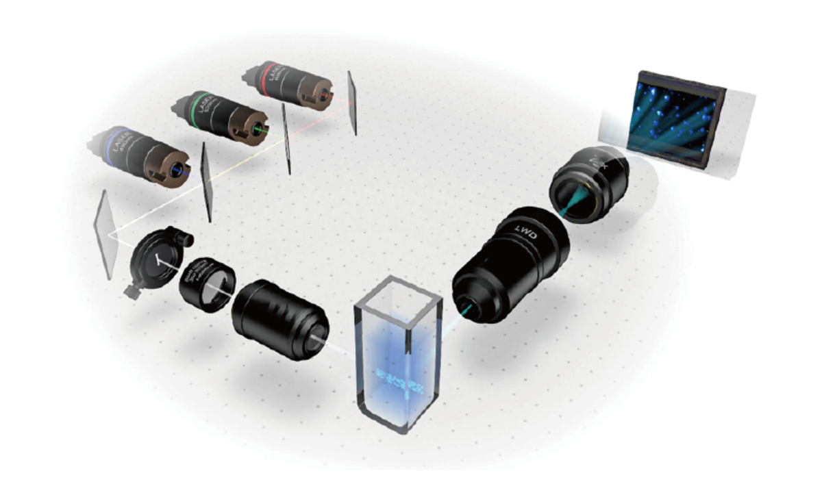
Simultaneous Multi-Laser Nanoparticle Tracking Analysis (NTA)
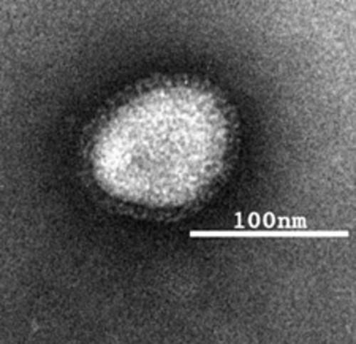
Exosomes? Virus? Nanoparticle? Use multiple lasers for complete, detailed analysis of all the particles in your sample.
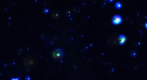
Exosomes, viruses, and nanoparticles all have wide size distributions which defeat traditional Nanoparticle Tracking Analysis (NTA) analyzers. The ViewSizer 3000 features simultaneous measurement with three lasers to collect the most accurate distribution and concentration information over a wide range of sizes within the same sample. Where the signal from a particle is too bright and saturates the detector from one laser, the software automatically uses data from a lower power laser to ensure the most accurate size and concentration information. On the other hand, when scattering from one laser is too weak for detection, the software uses data from a higher power laser to accurately track the particle.
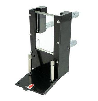
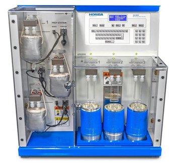
Cross contamination is a concern in all analyses. Simplified cleaning means thorough cleaning. The easy-to-remove sample cells can be dissassembled for rapid, thorough cleaning, which leads to better data.
Escape the limits of traditional NTA
Accurate and sensitive analysis without cross contamination
Scroll down and contact us for more information
The ViewSizer determines particle size and size distribution details by multi-laser nanoparticle tracking analysis (NTA). Multiple lasers enable analysis of a broad range of sizes in the same sample.
NTA can be used to count particles in the measurement volume. A patented method corrects for the effect of particle size on effective measurement volume.
The sample cell can be completely removed and dissassembled for complete cleaning. Disassembly, cleaning, and reassembly is faster than rinsing a flow cell. Furthermore, multiple cells can be used for faster throughput or assigned to various groups in a shared (core) facility.
| Biological characterization using protein crystal measurements | https://bioprocessintl.com/analytical/product-characterization/biological-characterization-using-protein-crystal-measurements/ |
| A lipase-independent pathway of lipid release and immune modulation by adipocytes | https://science.sciencemag.org/content/363/6430/989 |
| Application of a novel new multispectral nanoparticle tracking technique | https://iopscience.iop.org/article/10.1088/1361-6501/aab940/meta |
| Biophysical characterization of polydisperse liposomal adjuvant formulations | https://doi.org/10.1016/j.bbrc.2020.05.156 |
| Characterisation of particles in solution – a perspective on light scattering and comparative technologies | https://doi.org/10.1080/14686996.2018.1517587 |
| Cyclodextrin Reduces Intravenous Toxicity of a Model Compound | https://doi.org/10.1016/j.xphs.2019.01.004 |
| Development and anti-Candida evaluation of the vaginal delivery system of ... nanosuspension-loaded thermogel | https://doi.org/10.1080/1061186X.2018.1434660 |
| Electrochemical sensor based on F,N-doped carbon dots decorated laccase for detection of catechol | https://doi.org/10.1016/j.jelechem.2019.03.071 |
| Light scattering by pure water and seawater: the depolarization ratio and its variation with salinity | https://doi.org/10.1364/AO.58.000991 |
| Lipid Nanoparticle-Delivered Chemically Modified mRNA Restores Chloride Secretion in Cystic Fibrosis | https://doi.org/10.1016/j.ymthe.2018.05.014 |
| Mesenchymal Stromal Cell Bioreactor for Ex Vivo Reprogramming of Human Immune Cells | https://doi.org/10.1038/s41598-020-67039-w |
| Multifunctional Nanocomposites Based on Liposomes and Layered Double Hydroxides Conjugated with Glycylsarcosine for Efficient Topical Drug Delivery to the Posterior Segment of the Eye | https://doi.org/10.1021/acs.molpharmaceut.8b01136 |
| Particle size analysis of polydisperse liposome formulations with a novel multispectral advanced nanoparticle tracking technology | https://doi.org/10.1016/j.ijpharm.2019.06.013 |
| Review of nanoparticles in ultrapure water: definitions and current metrologies for detection and control | https://www.ovivowater.com/review-of-nanoparticles-in-ultrapure-water-definitions-and-current-metrologies-for-detection-and-control/ |
| Spark erosion as a high-throughput method for producing bimodal nanostructured 316L stainless steel powder | https://doi.org/10.1016/j.powtec.2018.01.012 |
| Synthesis and Characterization of EGFR-Targeted Immunoporphysomes | http://hdl.handle.net/1807/89548 |
| Synthesis of Ultrasmall Synthetic Melanin Nanoparticles by UV Irradiation in Acidic and Neutral Conditions | https://pubs.acs.org/doi/abs/10.1021/acsabm.9b00747 |
| Nanoparticle Tracking Analysis for the Quantification and Size Determination of Extracellular Vesicles | Protocol (jove.com) | https://doi.org/10.3791/62447 |
| Isolation and characterization of EV in Saliva of Children with Asthma | https://evcna.com/article/view/3962 |
| Spinal cord injury alters microRNA and CD81+ exosome levels in plasma extracellular nanoparticles with neuroinflammatory potential | https://doi.org/10.1016/j.bbi.2020.12.007 |
| Skeletal muscle tissue secretes more extracellular vesicles than white adipose tissue and myofibers are a major source ex vivo but not in vivo | https://doi.org/10.1101/2020.09.27.313932 |
| Human milk extracellular vesicle miRNA expression and associations with maternal characteristics in a population-based cohort from the Faroe Islands | https://www.nature.com/articles/s41598-021-84809-2 |
| Purification of Cas9 - RNA Complexes by Ultrafiltration | https://doi.org/10.1002/btpr.3104 |
| Assessment of Surface Glycan Diversity on Extracellular Vesicles by Lectin Microarray and Glycoengineering Strategies for Drug Delivery Applications | https://doi.org/10.1002/smtd.202100785 |
| Bioactive extracellular vesicles from a subset of endothelial progenitor cells rescue retinal ischemia and neurodegeneration | https://doi.org/10.1172/jci.insight.155928 |
| Circulating Extracellular Vesicles From Septic Mice Induce Brain Inflammation via Mirna Cargo | http://hdl.handle.net/10713/19055 |
Measurement Range | 10 nm to 15 μm |
|---|---|
Typical Sample Volume | 350 µL to 2.5 mL |
Typical Sample Concentration | 10^5 – 10^9 particles/mL |
Sample Temperature Range | 10° C to 50° C +/- 0.1° C |
Dimensions | 55 cm W x 66 cm D x 35 cm H |
Weight | 27 kg |
Operational Environment | 15° C to 30° C with < 85% RH |
Do you have any questions or requests? Use this form to contact our specialists.
Nanoparticle Analyzer
Laser Scattering Particle Size Distribution Analyzer
Static Image Analysis System Particle Size
Dynamic Image Analysis
Direct Imaging Particle Analyzer

Nanoparticle Analyzer
Centrifugal Nanoparticle Analyzer

Laser Scattering Particle Size Distribution Analyzer
Laser Scattering Particle Size Distribution Analyzer
Reticle / Mask Particle Detection System

Static Image Analysis System Particle Size
Reticle/Mask Particle Remover
BET Flowing Gas Surface Area Analyzers
BET Surface Area Analyzers