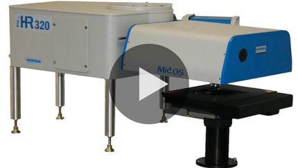
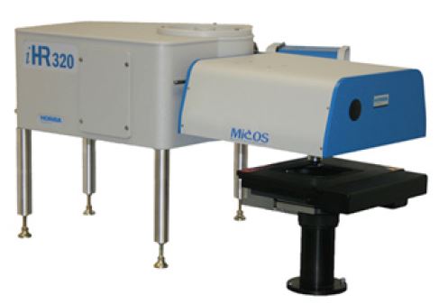
Bring the microscope to your spectrometer!
Using a standard microscope for luminescence characterization often means inefficient fiber-optic coupling to a spectrometer, and difficult access for many sample configurations, such as side-emitting devices, or upright cryostats. Nor do standard microscopes offer flexibility for coupling multiple lasers for photoluminescence excitation.
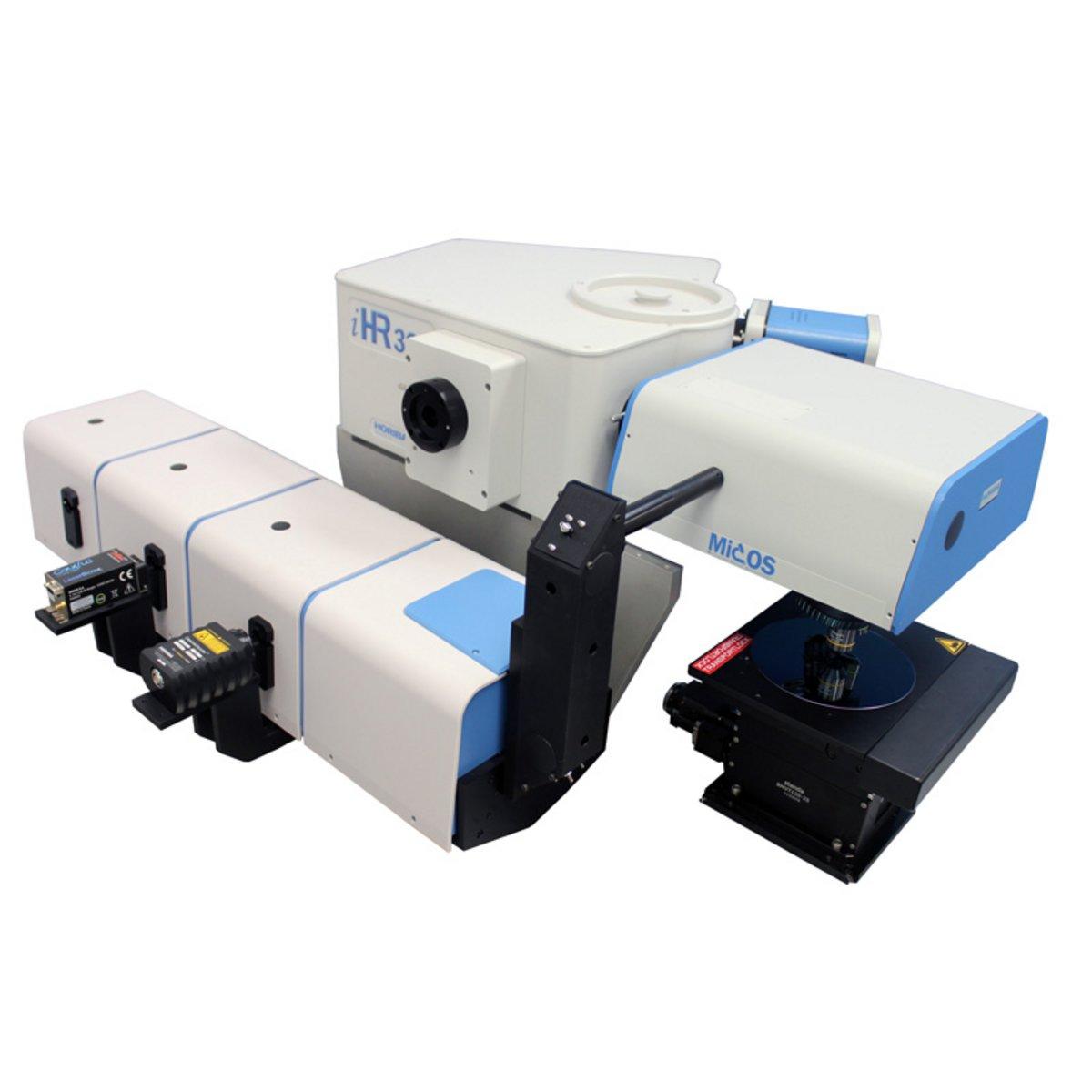
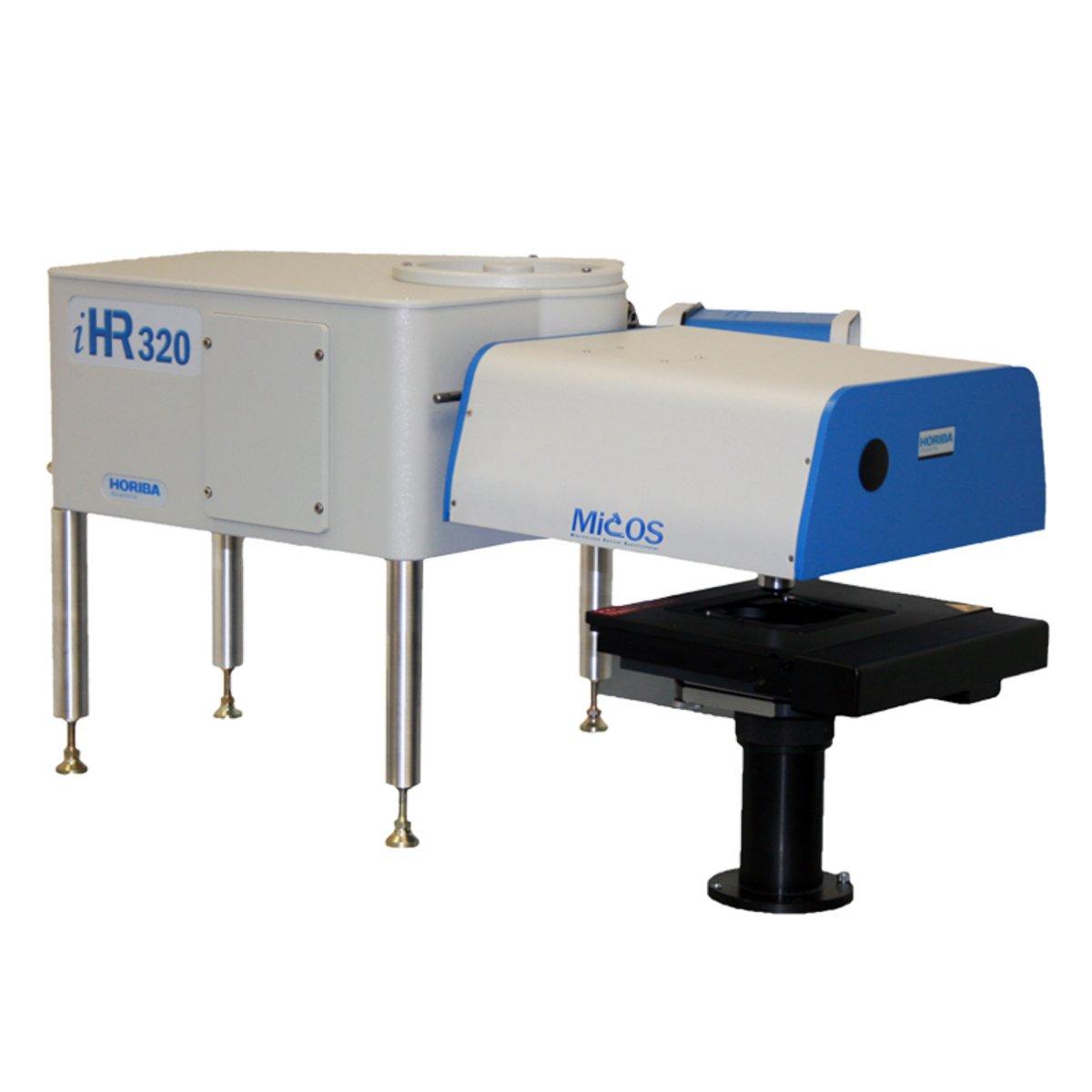
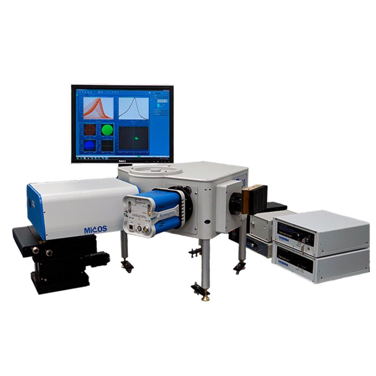
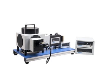
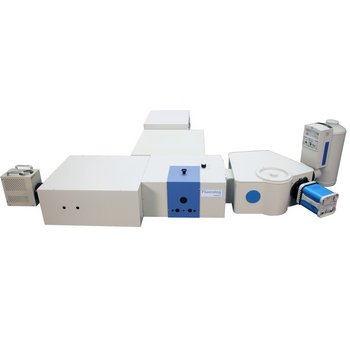
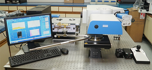
HORIBA Scientific’s MicOS merges microscopy and spectroscopy, to provide optimal coupling from the sample all the way to the detector. Available down- or side-looking configurations for side-emitting devices or upright cryostats give you flexible sample access. An optional, fully automated stage for mapping and sample-positioning is available. The MicOS offers a flexible platform for the use of multiple lasers for sample excitation. The system includes a vision camera so you always see what you are measuring.
Laser
The MicOS can be used with any combination of laser wavelengths, both single wavelength excitation and multiple wavelength excitations. The laser sources may either be fiber coupled to the MicOS input or assembled in a laser train to free space couple the light into the MicOS. Fiber coupling provides ultimate flexibility, and free space coupling provides the best possible spatial resolution with high magnification objectives.
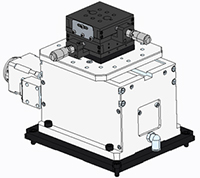
MicOS Microscope Head
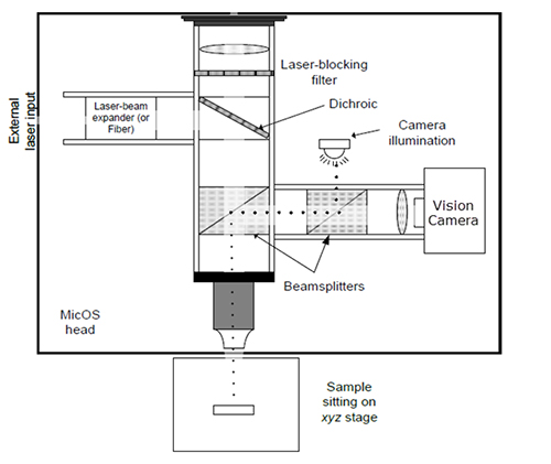
The MicOS microscope head includes all of the coupling optics for directing the laser to the sample and collecting the signal and coupling it to the spectrometer and detector. It also includes a white light source for illumination of the sample and a vision camera for viewing the sample. The optical schematic below shows the layout of the MicOS head.
Spectrometer

A range of spectrometers are available for integrating into your own Raman experiment, with focal lengths and resolutions to suit all requirements – ranging from 140 mm to 550 mm and above. Whether the application is for routine screening, fluorescence and PL mapping, or detailed high resolution Raman research, the spectrometer to match is available.
- High precision spectrometers with low stray light levels and options for high spectral resolution
- Gratings and sampling optics optimized for Raman
- Upgradeable spectrometer platform ready to expand to fluorescence, PL, and optical spectroscopy
Click here to view all of the available spectrometers.
Detector
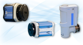
A range of detectors are available for integrating into your own Raman experiment, with different chip architectures and various degrees of cooling to suit all requirements. Whether the application is for rapid measurements or very weak Raman scattering samples, there is a detector to match.
- High sensitivity CCD detectors for UV-VIS
- Linear InGaAs arrays for near-IR response
- Deep cooling for low dark current (thermoelectric or LN2 cooling)
Click here to view all of the available detectors.
Software
LabSpec 6 Spectroscopy Software
The LabSpec6 spectral software suite used on all the HORIBA Jobin Yvon analytical and research Raman spectrometer systems is now also available for modular Raman systems. It has been designed and written as a dedicated Raman spectroscopy package and offers many powerful capabilities not found in a basic spectroscopy software.
Accessories
Cryostats
Depending on the MicOS configuration (down- or side- looking), various models of cryostats may be employed. An example is given below, however the MicOS system is customizable to a wide variety of cryostat types and manufacturers.
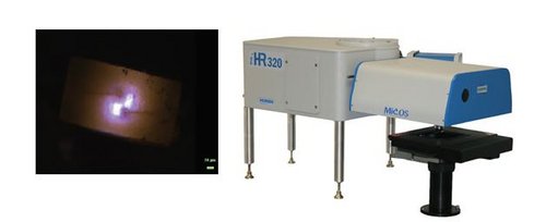
Sample Stages
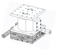
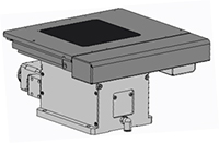
The manual XYZ stage is ideal for low budget applications where mapping is not critical, but precise positioning of the sample under test is crucial.
A variety of sample stages are available, both manual and motorized. Schematics are shown below for various options.
The manual XYZ stage is ideal for low budget applications where mapping is not critical, but precise positioning of the sample under test is crucial.
The manual XY, motorized Z stage is ideal for applications where real time mapping is not required, but fine control over the focus (Z) is desired.
The motorized XYZ stage is required for any applications where real time mapping of the sample under test is desired. The controller integrates seamlessly with LabSpec software, providing a complete, turn-key system.
Optical Breadboard
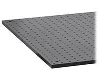
For users who do not have access to an optical table, a 2’ x 4’ breadboard is available so that the MicOS can be set up on any standard table.
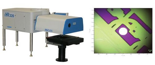
Wafer Holder
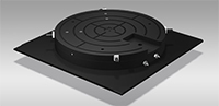
The microscope stage can be equipped with a wafer holder for mapping of large areas. A rotating 4” wafer holder is standard, however larger wafer sizes can be accommodated. Contact the factory for details.
 This product has been discontinued and is no longer available. This page is provided for informational purposes only.