Wear metals and additive Elements Analysis.

 This product has been discontinued and is no longer available. You can still access this page for informational service purposes.
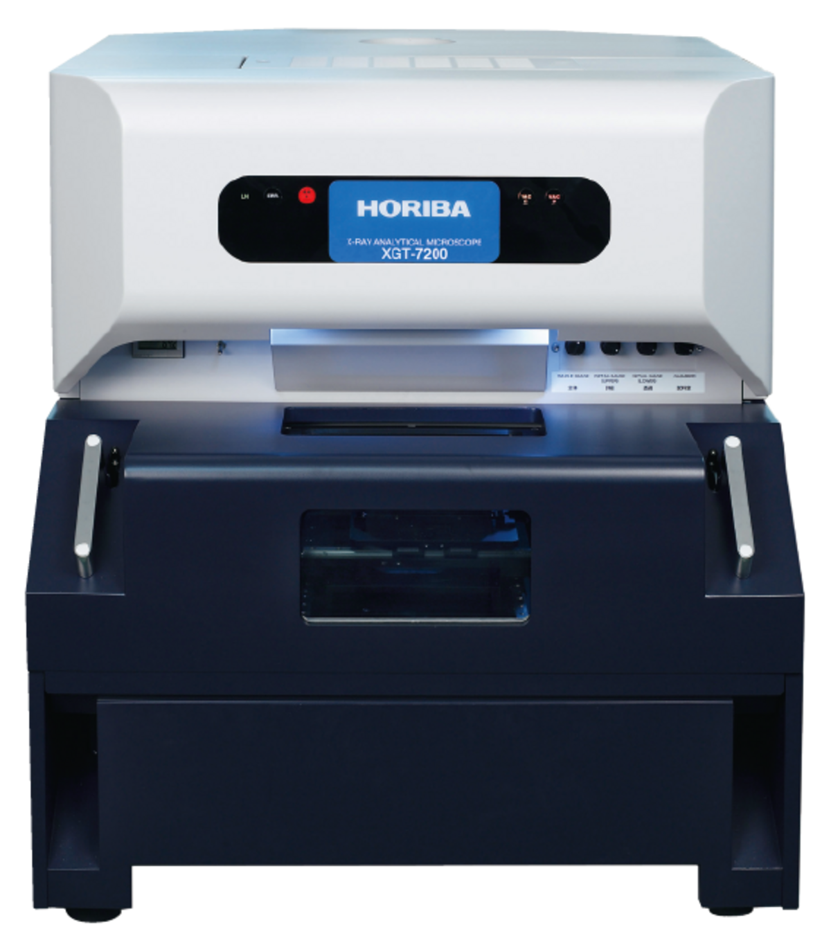
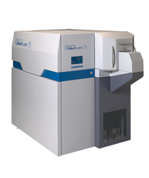
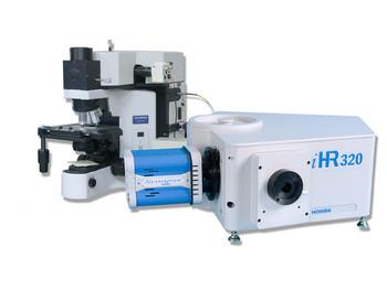
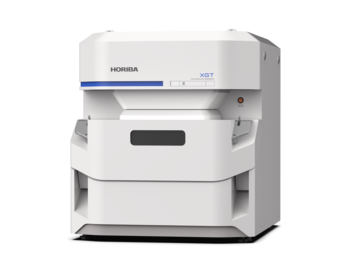
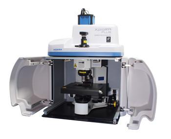
X-ray Analytical Microscope
The XGT-7200 represents a completely new generation of XRF microscope, and leads the way to a new era of science. It offers a seamless merger between optical observation and elemental analysis functions, revolutionizing the world of micro-analysis and establishing micro-XRF as a routine tool for the research and analytical scientist.
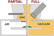
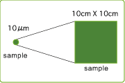
Unique hardware features ensure the system offers versatility and flexibility for every measurement. A choice of two software controlled x-ray guide tubes with diameters ranging from a unique 10 µm through to 1.2 mm allow conditions to be optimised for a range of measurements, including both micro and macro. Similarly, with the unique Dual Vacuum Modes it is possible to switch within seconds between a high sensitivity full vacuum mode and a versatile localised vacuum mode. The latter maintains samples at atmospheric pressure whilst retaining sensitivity to all elements from sodium to uranium.
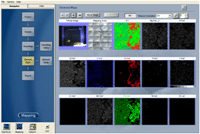
In any configuration the intelligent combination of optical cameras ensure that the precise analysis position can be quickly and simply located.
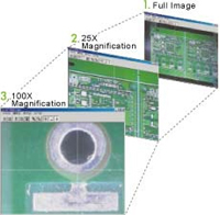
Quickly proceed from a view of the entire sample to the selection of the analysis potision.
Intuitive "click and move" images from low magnification and high magnification cameras within the sample chamber, allow the analysis position or mapping area to be defined withing seconds.
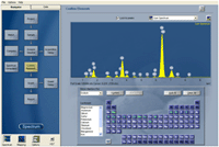
Single Point and Multi-point Analysis
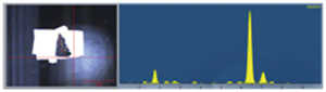
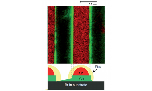
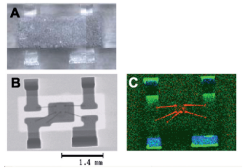
Single point and automated multi-point analyses allow high quality spectra to be acquired from either a single position, or from a number of user defined points across the sample. Element peaks are automatically located and labelled, and quantitative analysis down to ppm levels can be carried out using the fundamental parameters method (FPM), FPM with single standard, and full standard sample calibration.Thickness calculations can also be made on nm and µm thick multi-layered structures.
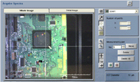
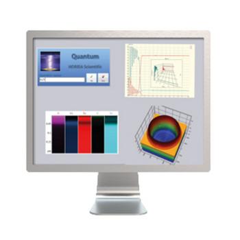
Hyperspectral Mapping Analysis
The SmartMap imaging software records a full EDXRF spectrum at each and every pixel of the element image, enabling post-acquisition element image generation and comparison, and spectrum generation from user defined regions in the image with subsequent qualitative and quantitative characterisation.
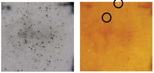
Transmitted X-ray imaging provides additional insight into a sample’s structure, allowing features invisible by eye to become immediately apparent.
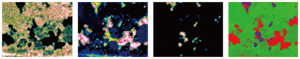
Element image display
Element images to be displayed can be selected during and after acquisition. Images can aslo be generated from user defined spectral windows.
RGB Composite Image Generation
Overlay elemental images to allow easy comparison of element distribution
Spectrum Generation
Generate the average spectrum from a user defined region within an image.
Line Analysis
Display multiple element intensity profiles across a defined region of an image.
Data Export
Element images can be exported in data or image formats. The entire hyperspectral datacube can be exported in RAW format.
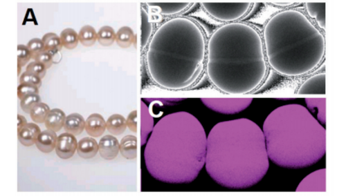
The unique features of the XGT-7200 have seen this innovative micro-XRF analyser widely embraced for a range of applications, including electronics, engine wear analysis, forensic science, geology, mineralogy, pharmaceutics, museums, metallurgy, biology, medicine and archaeology.
The flexible XGT-7200 micro-XRF system covers everything from macro analysis, for a general survey of a wide area, to the inspection of a specific micro area, with simultaneous XRF and transmission imaging. Its many features ensure high performance analysis with easy operation.


XGT-7200 Specification
Elements: Na to U
X-ray tube: Rh target / Tube voltage 50kV / Tube current 1mA
Fluorescent X-ray detector: Peltier cooled Silicon Drift Detector (SDD)
Transmitted X-ray detector: Nai(Tl) scintilltor
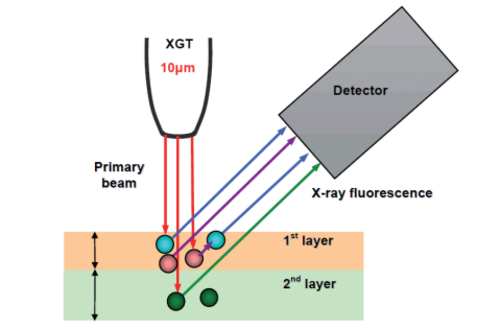
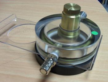
X-ray guide tube: Mono capillary 10μm / 100μm with no filter
Optical image: Full sample optical image and coaxial magnification image
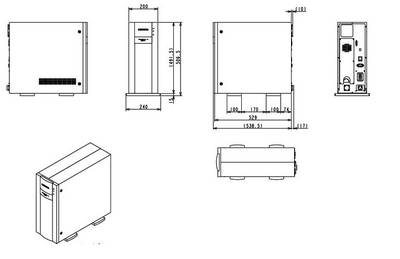
Sample stage size: XY : 100mm×100mm
Sample chamber: Vacuum chamber model/ Max 300mm×300mm×80mm in vacuum
Signal processing: Digital pulse processor ( INCA unit )
Qualitative analysis: Auto identification / KLM marker / Peak search / Compare spectrum
Quantitative analysis: Non standard FPM / Standard FPM / Standard file matching FPM /Calibration curve / Multi layer FPM (Thickness gage) /Multi point analysis (Max5000) / Multi point result send to Excel / XGT-5000 spectrum view
Mapping function: Transmitted X-ray image / Elemental image / Spectrum mapping / Rectangle mapping / Generate spectrum / RGB composition / Scale maker / Line analysis
Other function: Possible to start the XGT-5200 software simultaneously.

Do you have any questions or requests? Use this form to contact our specialists.
X-Ray Fluorescence Analyzer
X-Ray Fluorescence Analyzer
High resolution, high sensitivity and high stability ICP-OES
Affordable high performance ICP-OES
X-ray Analytical Microscope (Micro-XRF)
Air Pollution Monitor
Ambient Dust Monitor
Ambient Air Quality Monitoring System

Spectroscopic Ellipsometer for Simple Thin Film Measurement
Customize your instrument

Intuitive Auto-Soft Interface for the Auto SE and Smart SE
Hybrid Chemical and Video Image Display
A Platform for HORIBA Scientific Ellipsometers
Pulsed-RF Glow Discharge Optical Emission Spectrometer
Accessories for samples with various shapes, sizes and properties
Quantum and Image
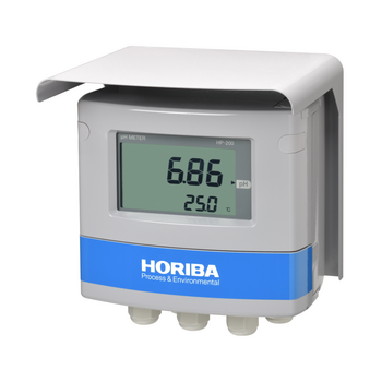
Field-installation type water quality measuring instruments

ICP Software

Complete your ICP-OES Spectrometer for your specific needs
Emphasize Raman and Optical Images

For your specific application
AFM-Raman for physical and chemical imaging

Raman Spectroscope - Automated Imaging Microscope

Real-time and Direct Correlative Nanoscopy
LabSpec 6 is a validated software

Camera Endpoint Monitor based on Real Time Laser Interferometry
X-Ray Fluorescence Analyzer
X-Ray Fluorescence Analyzer
Recall settings, and automate processes

Solid Particle Counting System
Data analysis for complex data sets

On-board Emissions Measurement System
Fast and easy Raman acquisition

Manual label-free molecular interaction analysis machine Flexible Research Platform
Fiber coupled microscope
Customize with VBS

Powerful and Cost Effective Spectroscopic Ellipsometer
PTFE Filter/TFH membrane

High resolution, high sensitivity and high stability ICP-OES

Affordable high performance ICP-OES
Password protected user access control

Spectroscopic Ellipsometer from FUV to NIR: 190 to 2100 nm
X-ray Analytical Microscope (Micro-XRF)
X-ray Analytical Microscope
with a Super Large Chamber

AFM-Raman for Physical and Chemical imaging
MicroRaman Spectrometer - Confocal Raman Microscope
X-Ray Fluorescence Analyzer
X-Ray Fluorescence Analyzer
X-ray Fluorescence Sulfur/Chlorine-in-oil Analyzer
X-ray Fluorescence Sulfur-in-Oil Analyzer
X-ray Fluorescence Sulfur-in-Oil Analyzers
X-ray Fluorescence Sulfur-in-Oil Analyzer
X-ray Fluorescence Sulfur-in-Oil Analyzer
Energy Dispersive X-ray Fluorescence Analyzer
X-ray Analytical Microscope (Micro-XRF)
X-ray Analytical Microscope
with a Super Large Chamber