LabRAM Soleil Raman Microscope presentation

LabRAM Soleil™: Your Application at a Glance

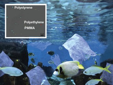
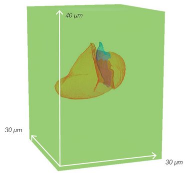
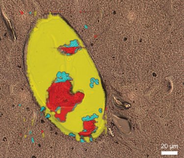
From Material Sciences to Life Sciences, from Environment to Earth Sciences, Raman microscopy can now easily reach unknown worlds.
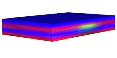
The LabRAM Soleil™ and its software have been developed to allow the characterization of all types of samples. Whether they are flat, rough or particulate, LabRAM Soleil™ characterizes them at high speed: up to 100 times faster than before. Thanks to its large sample compartment and the QScan™ system, even large and heavy samples can be characterized. Browse through these applications, and if you can't find yours, contact our experts who will be happy to test your samples.

 University of La SAPIENZA, Rome, Italy - Claudia Fasolato, Research Scientist:
The LabRAM Soleil is so compact, well-illuminated and versatile that you can measure any kind of sample with it!
It’s the ideal tool for our physics and biophysics research group, where we’re working on a wide variety of applications from nano-objects to perovskites and biological samples.

2D Materials

Application in 2D materials: Correlated PL and ULF Raman microscopies of WS2 sample

Correlated PL and ULF Raman microscopies of WS2 sample
2D materials are state of the art in nano- and opto-electronics. Characterizing their structural properties with a non-destructive approach at the micron scale is an important point.


Pharmaceutical tablet

Pharmaceutical tablet compounds distribution based on Raman spectral mapping from low to high concentrations (0.1% to 100%).
On the final step of the formulation process, pharmaceutical and cosmetic industries have to control their products to check the compounds distribution in the final product.

Download application note: Pharmaceutical compounds distribution and cosmetic products analyzed by confocal Raman spectroscopy

Download application note: Polymorphisms characterization: when Raman microscopy supports the pharmaceutical industry

Download application note: Morphological and chemical characterization of pharmaceutical formulations

 VIBRA-SANTÉ HUB, Center for Interdisciplinary Research on Medicines, Liege, Belgium – Eric Ziemons, Director:
With a multimodal Raman microscope as easy to set up as the LabRAM Soleil, we were quickly up to speed. We were able to study our pharmaceutical samples safely and without risk of contamination as it has Class 1 capability built in. It’s the ideal tool for the analyst of the future! 

Multilayer polymer

Multilayer polymers with buried artefacts mapped with QScan
Raman microscopy is an excellent tool to answer the polymer questions. It can be used to characterize raw materials, monitor polymerization process, investigate orientation and crystallization changes, and also to control the quality and traceability of genuine products by understanding defects and compounds distribution.

Download application note: Characterization of protective mask fibers by Raman microscopy

Download application note: Raman Microscopy Applied to Polymer Characterization: An Overview

Microplastics

Microplastics sorting down at the μm scale - PMMA (yellow), Polystyrene (blue), Polyethylene (red)
In a world where climate changes faster and faster, controlling pollutants becomes crucial. Raman microscopy is now the most effective technique to investigate morphology and composition of a large panel of particles like microplastics and aeroallergens.

Download application note: Analysis of microplastics in hand sanitizers using ParticleFinder™

Download our e-book: Microplastics

Download application note: Microplastics

 IFREMER, Brest, France – Maria El Rakwe, Research scientist:
We really like the LabRAM Soleil’s optimized design, which speeds up analysis of microplastics. It has helped us to improve our profitability, no matter what kind of environmental sample we’re looking at (notably water). 

Confocal 3D imaging of an inclusion

2.5 M pixels image of high definition image of a fluid inclusion in a quartz matrice
Confocal Raman microscopy is used in geochemistry and petrology to analyze components trapped in fluid, gaseous or solid inclusions in minerals. Raman can so shed some light on the origins of the universe.

Cellular imagery

SWIFT High Resolution Raman map of a mouse liver cell (500ms per point, 500nm step resolution, 70 000 points)
Studying how cells work is of fundamental importance in cellular and tissue biology. Raman spectra offer a wealth of information useful to biochemists, such as cell phenotype by analyzing nucleic acids, carbohydrate, lipid and protein content, etc.

Request for Information

Do you have any questions or requests? Use this form to contact our specialists.

* These fields are mandatory.

Corporate