La OCT (Tomografía de Coherencia Óptica) es una técnica de imagen que utiliza interferencias lumínicas para capturar imágenes de la estructura interna de una muestra con alta resolución y alta velocidad. En los últimos años, se ha utilizado principalmente como equipos de examen del ojo ocular en oftalmología. Además, la capacidad de imagen en tiempo real sin contacto no invasiva la hace aplicable en diversos campos, como la visualización de la estructura vascular interna de la piel para fabricantes de cosméticos, la observación celular para el mercado biotecnológico y la medición de formas de piezas metalúrgicas. Puede personalizarse para diferentes rangos de longitudes de onda, desde luz visible hasta infrarrojo cercano, dependiendo de cada aplicación.

Espectrómetro compacto para OCT
OCT significa Tomografía de Coherencia Óptica, que es una técnica de imagen 3D que permite mediciones sin contacto y no destructivas de alta resolución de la estructura interna de una muestra mediante la interferencia de la luz. Con una resolución de imagen de unos pocos micrómetros, la OCT puede medir estructuras tanto en superficie como en profundidad hasta varios milímetros. La OCT se ha desarrollado principalmente en el campo de la oftalmología. También se emplea en diversos campos, incluyendo inspecciones industriales en línea, como el examen de vasos sanguíneos, piel y muestras celulares.
SD-OCT (OCT en el dominio espectral) es una técnica que consiste en dirigir luz de banda ancha hacia el objetivo y obtener información de profundidad mediante la transformación de Fourier de la longitud de onda de la luz de interferencia reflejada por el objetivo.
Configuración básica (dominio del tiempo)
OCT utiliza fuentes de luz de baja coherencia como los diodos superluminiscentes (SLD). El haz desde la fuente se divide mediante un divisor de haz, con una parte dirigida hacia el objetivo y la otra hacia un espejo de referencia. La luz que entra en el objetivo se refleja o retrodispersa debido a diferencias en los índices de refracción y los cuerpos de dispersión dentro del objetivo. La luz de la muestra y del espejo de referencia se solapan. Las intensidades de la muestra y del espejo de referencia se solapan. Los haces de la muestra y del espejo de referencia solo se interfieren cuando los tiempos de viaje de la luz que regresa de la muestra y del espejo de referencia son aproximadamente iguales. La SLD es una fuente de luz de baja coherencia con un amplio espectro, similar a un LED, y alta luminosidad, similar a un diodo láser (LD).
Para medir la estructura interna, es necesario mover intencionadamente el espejo de referencia a lo largo del eje óptico. Al variar el tiempo de viaje de la luz desde el espejo de referencia mientras se mide la intensidad de la luz en cada posición, se produce interferencia en diferentes posiciones del espejo de referencia en movimiento debido a las diferencias en los tiempos de viaje de la luz que regresa de la estructura interna del objetivo. La intensidad de luz obtenida, representada en función del movimiento del espejo de referencia a lo largo del eje x, proporciona información sobre la estructura interna de la muestra. Además, escaneando en la dirección xy usando dispositivos como espejos galvanométricos, se pueden obtener imágenes 2D o 3D.
OCT en dominio de Fourier
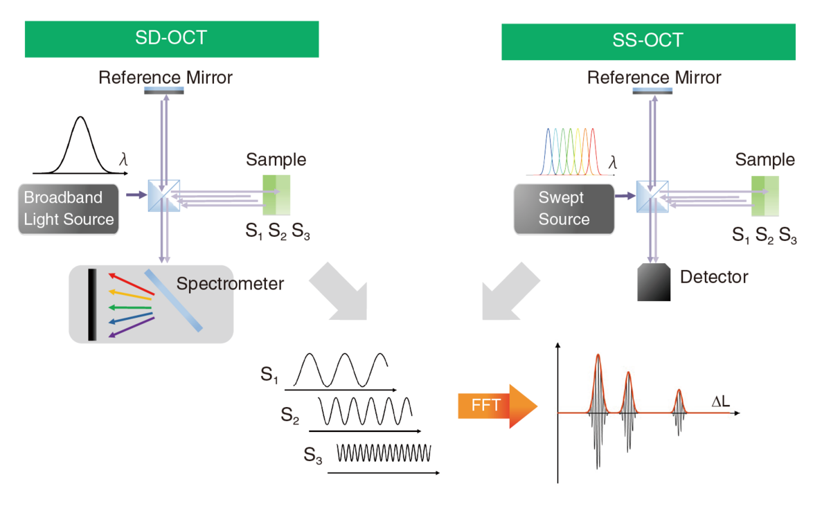
El OCT en dominio de Fourier (FD-OCT) es una técnica que mejora aún más la velocidad y sensibilidad en comparación con las OCT en dominio temporal. En lugar de mover el espejo de referencia, utiliza un espectrómetro o una fuente de luz barrida por longitud de onda, respectivamente, para medir la intensidad de las señales de interferencia a lo largo del rango de longitud de onda y obtener un espectro de interferencia. Dado que las frecuencias del espectro de interferencia obtenido corresponden a las posiciones en profundidad de varios límites dentro de la muestra, se pueden lograr mediciones similares a la OCT en el dominio temporal mediante la transformada de Fourier de estos datos. Esto permite la medición simultánea de todas las posiciones en profundidad dentro de la muestra sin necesidad de piezas mecánicas móviles, resultando en mediciones de alta velocidad y altamente sensibles en tiempo real sin ruido innecesario.
FD-OCT puede dividirse en dos categorías principales según sus configuraciones: OCT en dominio espectral (SD-OCT) y OCT de fuente barrida (SS-OCT). SD-OCT utiliza una fuente de luz de banda ancha y un espectrómetro como detector para adquirir simultáneamente espectros de interferencia. Por otro lado, SS-OCT mide la intensidad de la luz en cada longitud de onda barriendo la longitud de onda de la fuente de luz y luego disponiéndolas temporalmente para obtener espectros de interferencia.
Estas dos técnicas difieren en resolución, profundidad de medición, estabilidad, velocidad y facilidad de adquisición de componentes. Se eligen en función de propósitos y aplicaciones específicas. Además, los avances recientes han llevado al desarrollo de la OCT multifuncional, que utiliza estas técnicas no solo para capturar información estructural del objetivo, sino también para obtener propiedades de materiales y otra información valiosa.
Selección de longitud de onda
Para SD-OCT, el espectrómetro es uno de los componentes clave que debe personalizarse según la muestra de medición. Primero, tienes que elegir el rango de longitudes de onda como rango visible, rango de 800 nm, rango de 1000 nm, rango de 1300 nm, etc. La elección depende de factores como las características de absorción y dispersión de la muestra, así como de la disponibilidad de los componentes. Por ejemplo, en exámenes retinianos, a menudo se prefieren longitudes de onda alrededor de 800 nm o 1000 nm para minimizar la influencia de la absorción de agua.
A continuación, considera la resolución de profundidad\(\delta_z\)y el rango de profundidad\(Z_{max}\). Estos se determinan principalmente por la fuente de luz y el espectrómetro. Dando la longitud de onda central de la fuente de luz como\(\lambda_c\), el ancho completo a la mitad del máximo (FWHM) de la fuente de luz como\(\Delta\lambda_L\), la resolución en píxeles del espectrómetro como\(\delta\lambda_s\)(rango de longitudes de onda del espectrómetro\(\Delta\lambda_s\)dividido por el número de píxeles del detector\(p\)) y el índice de refracción de la muestra como\(n\), pueden expresarse de la siguiente manera:
\(\delta_z=n/2In2\) * \(\lambda_c^2/\Delta\lambda_L\)
\(Z_{max}=1/4n\) * \(\lambda_c^2/\delta\lambda_s\)
Los dos diagramas siguientes muestran la resolución de profundidad y el rango de profundidad para cada longitud de onda central. Para lograr una mejor resolución de profundidad, se requiere una fuente de luz de mayor ancho de banda. Sin embargo, un espectrómetro diseñado para dicha fuente de luz suele tener una mayor resolución de píxeles, lo que resulta en un rango de profundidad más estrecho. Como resultado, existe una relación de compromiso entre la resolución de profundidad y el rango de profundidad.
Un espectrómetro que cubra todo el rango de longitudes de onda de la fuente de luz no es suficiente. Por ejemplo, al usar una fuente de luz con un ancho de banda de 800-900 nm, necesitarías un espectrómetro que cubra un ancho de banda similar. Usar un espectrómetro que cubra un rango más amplio, como 700-1000 nm con el mismo sensor, limitaría el rango de profundidad de aproximadamente 3,7 mm a solo 1,2 mm. Para maximizar el rendimiento, es fundamental contar con un espectrómetro personalizado para que coincida con las características de la fuente de luz utilizada.
Sensibilidad
La sensibilidad de la señal de interferencia disminuye con la profundidad, principalmente debido a una menor reproducibilidad del espectro de interferencia en sitios más profundos. A medida que aumenta la profundidad, el espectro de interferencia contiene frecuencias más altas. Sin embargo, dado que el tamaño del píxel de la cámara y el tamaño del punto de recogida del espectrómetro tienen dimensiones finitas, la reproducibilidad empeora con el aumento de la profundidad, lo que resulta en una menor intensidad de la señal. Esta relación entre sensibilidad\(R(z)\)y profundidad\(z\)puede expresarse de la siguiente manera:
\(R(z)=\frac{\mathrm \sin^2 \begin{bmatrix} D(z)\end{bmatrix}}{\mathrm D(z)^2}・exp \begin{bmatrix} - \frac{w^2}{2In2}D(z)^2\end{bmatrix}\)
\(w\)representa la relación de resolución en longitud de onda\(\delta\lambda_R\)a resolución de píxeles\(\delta\lambda_s\). La atenuación de la sensibilidad calculada por esta ecuación, para dos tamaños de puntos de haz enfocados (= resolución de longitud de onda, x1 y x2 en comparación con el tamaño del píxel) se muestra en la figura siguiente. Es evidente que el impacto del rendimiento del enfoque de haz en la sensibilidad es considerable. Los espectrómetros necesarios para la OCT difieren de los espectrómetros típicos y deben diseñarse de modo que el tamaño del haz enfocado en el detector sea muy pequeño, por ejemplo, menor que el tamaño del píxel.
Tiene alguna pregunta o solicitud? Utilice este formulario para ponerse en contacto con nuestros especialistas.
Espectrómetro compacto para OCT