Size:
6.6
MB
ParticleFinder™ for Raman Spectrometer Brochure
Automated Particle Measurement, Identification and Classification using Raman Analysis
ParticleFinder™ provides an integrated and customizable workflow for particle analysis.
It automatically detects particles, even on complex and patterned holders, analyzes them for size and shape descriptors, performs chemical characterization using Raman microscopy, and identifies them with a HQI (Hit Quality Index).
Offering fully automated multi-sample analysis, ParticleFinder generates comprehensive reports based on customizable templates, ensuring routine-ready microparticle analysis.

Check out our LabSpec 6 Video Resource Center to fully master your LabSpec 6 spectroscopy software!
ParticleFinder is compatible with any HORIBA Raman spectrometer equipped with LabSpec 6 software, a video camera and a motorized XY sample stage. With these requirements met, ParticleFinder’s Raman analysis can fully exploit the unique capabilities of HORIBA’s Raman systems to ensure the most appropriate chemical interrogation. This can range from routine identification of common particles and contaminants, to advanced characterization of polymorphism/phase, photoluminescence and stress/strain.
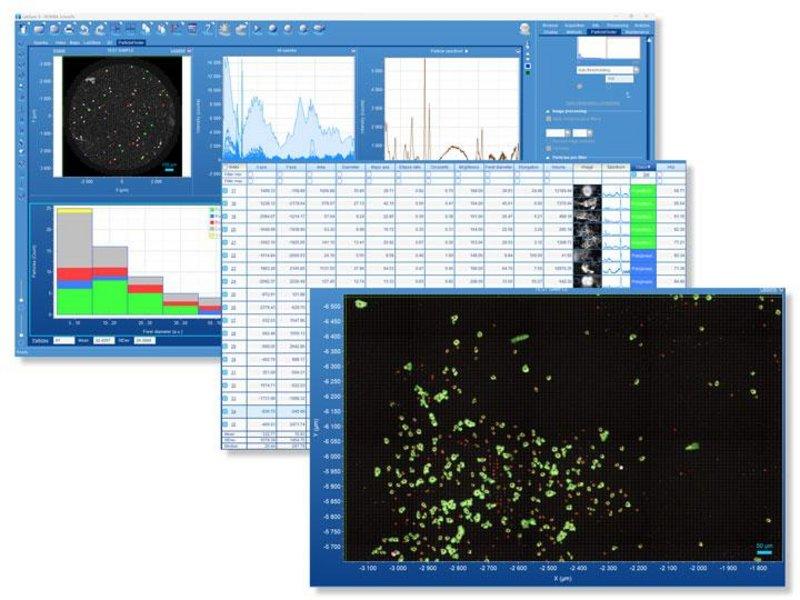
LabSpec 6 screenshot: Particle detection overlaid on white light optical image, with morphological and chemical characterization
The workflow for ParticleFinder is extremely simple and intuitive. The module is an integrated part of the powerful LabSpec 6 software, and is fully linked to related modules for data acquisition, processing, analysis, spectral identification and display.
Simple processing of the sample optical image quickly locates the particles, creating a binary image which clearly highlights the particle positions. Full morphological filtering tools are included to ensure truly meaningful location. The optical image can cover any user-specified area, using the integrated video montage/mosaic tool, which allows millimeter or centimeter areas to be covered.
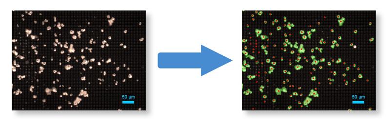
ParticleFinder automatically locates particles even on complex or patterned holders
Once the particles are located, particle shape and size parameters are calculated for each particle, including a statistical summary. Histograms of the parameters (including area, perimeter, axes, ellipse ratio, circularity, and brightness) are available for easy visualization.
ParticleFinder calculates the shape and size of located particles and generates a statistical summary
Initially, all located particles are set for Raman analysis. However, they can be filtered according to the statistics results, allowing only particles which correspond to a specific size/shape to be analyzed. For example, only particles within a certain size range can be marked for Raman characterization.
Alternatively, ParticleFinder enables sample randomization to achieve statistically significant sample analysis while reducing overall analysis time.
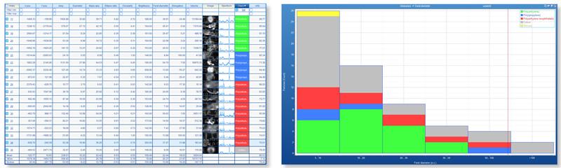
The full capabilities of the Raman instrument can be utilized for the Raman acquisition stage, including choice of laser wavelength, spectral resolution, confocality and mapping. Such configurable experiments ensure that optimized results can be obtained from varying sample sets with ease. The Raman spectra are displayed in a table together with the statistical parameters.
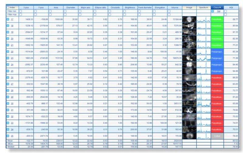
ParticleFinder displays Raman spectra together with the statistical parameters
Once the Raman data has been acquired, it is automatically linked to the individual particles in the binary image. The spectra are now available to be processed, analyzed and displayed according to the full powerful functionality of LabSpec 6.
Spectra can be automatically identified using the fully integrated IDFinder app, particle-by-particle, with HQI (Hit Quality Index).
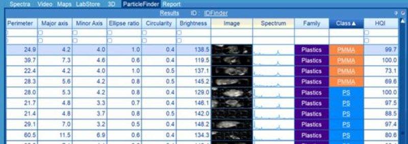
ParticleFinder automatically identifies spectra using the fully integrated IDFinder app, particle-by-particle, with HQI
Alternatively, the fully integrated Multivariate Analysis module can be used for automatic clustering and decomposition of the spectra.
ParticleFinder generates statistics based on various morphological or chemical information, offering fully customizable histograms, scatter plots, or tables.
The spectral identification results are presented in the statistical table and directly overlaid on the video image of your sample, where each color corresponds to a different component.
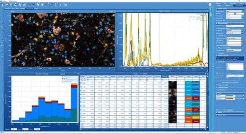
ParticleFinder has a user-friendly interface showing particle identification images, spectra and statistics results in a single screen
ParticleFinder enables the creation of comprehensive, multi-page report templates that can include histograms, images, tables, scatter plots, and more, along with company and sample-related information. These report templates are then utilized to generate automated reports for each analyzed sample.
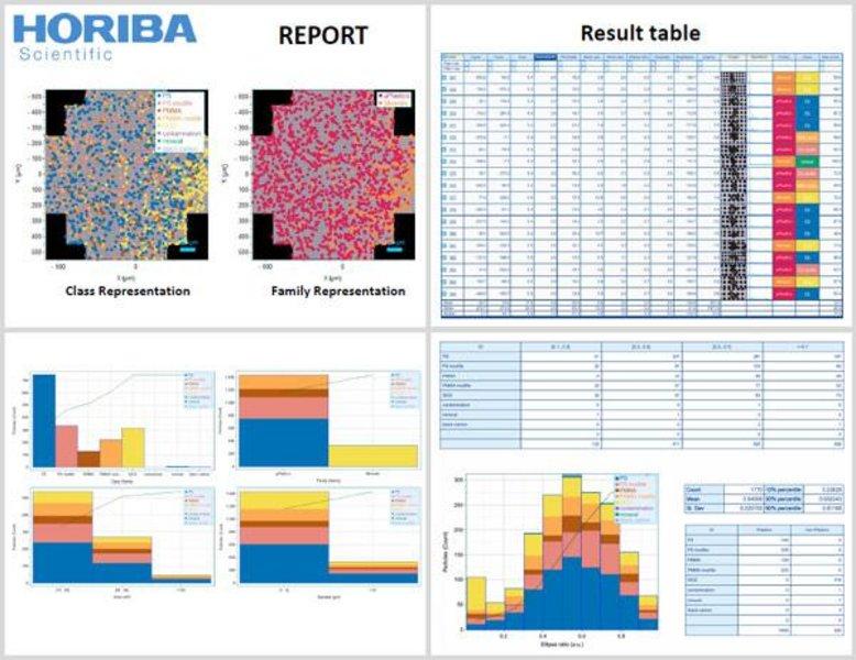
ParticleFinder enables the creation of comprehensive, multi-page reports
Set up all of your measurements for multiple samples at one time, using the HORIBA sample holders, or any multi-sample support.
ParticleFinder supports any design of sample holders fitting on the motorized stage, and provides fully automated measurements for 24/7 operation.
ParticleFinder performs simple, automated, and precise particle sample analysis, thus making research methods suitable for deployment in process labs.
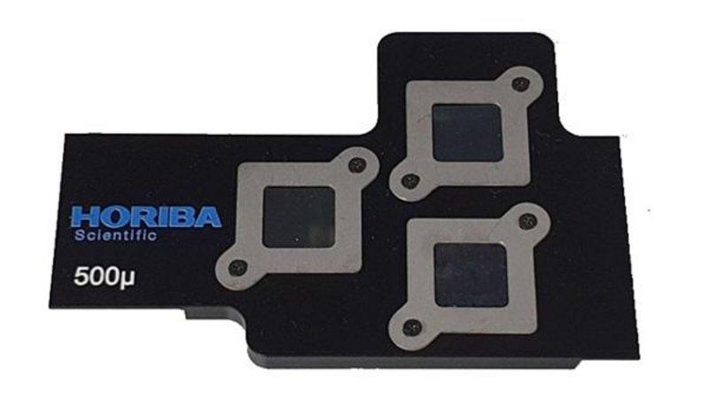
HORIBA 10x10mm Silicon filter multi-sample holder
The ParticleFinder module for Raman spectrometers is one part of HORIBA’s expertise in particle analysis. HORIBA offers powerful, hassle-free tools for particle size, particle shape, zeta potential, and surface area analysis. Measurable particle size range starts at below 1 nanometer and goes up to 30 millimeters, at concentrations ranging from 1 ppm to 50 vol% with shape determination available starting at 1 micrometer. A range of technologies are employed, including laser diffraction, dynamic light scattering, and dynamic and static image analysis.
Do you have any questions or requests? Use this form to contact our specialists.
Particle Disperser
