PDF
1.17
MB

Raman Resonance Raman Spectroscopy of Enzymes

Molecular structure of PNA photolyase binding in close proximity to FAD cofactor.
Raman and resonance Raman spectroscopy have proven to be important research tools to investigate structure-function relationships in enzymes.
One such enzyme is DNA photolyase, which is a blue-light photoreceptor and uses flavin adenine dinucleotide in a light-driven, electron-transfer mechanism to repair cyclobutane pyrimidine dimers of DNA. In order to understand the intricate interactions between the FAD cofactor and its protein environment better, it is essential that the assignments of the vibrational modes are well understood.
In photolyase, the cyclobutane pyrimidine dimer of DNA binds in close proximity of the FAD cofactor, and one of its carbonyl groups in nearly van der Waals contact with the C(8)- methyl group of the redox-active isoalloxazine ring of FAD. However, the vibrational modes that are associated with the C(8)-methyl group and could report on important enzyme-DNA interactions, are yet unknown. A fully integrated HORIBA Scientific spectroscopy system was used to determine the vibrational modes of flavin that are sensitive to motion of the C(8)-methyl group.
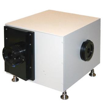
Short Focal Length Triple Grating Imaging Spectrographs
Dual Mode Analog/Photon Counting PMT
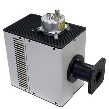
Confocal Raman & High-Resolution Spectrometer
HORIBA제품의 자세한 정보를 원하시면, 아래의 양식에 내용을 입력을 부탁드립니다.