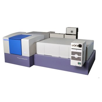
Confocal Laser Scanning FLIM
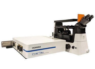
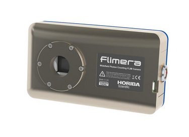
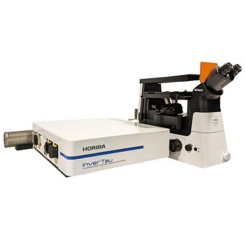
InverTau™ is our new confocal laser scanning FLIM platform. InverTau is fully software controlled and designed to fit on the side port of an inverted fluorescence microscope. You can build your own microscope system around the InverTau platform, or buy a complete microscope system from HORIBA. InverTau can be enhanced to acquire real-time wide field video rate FLIM up to 30 frames per second with the addition of FLIMera. Versatile solutions are always available with local support.
- Affordable laser scanning unit for FLIM studies
- Fully computer-controlled confocal optics, with the intuitive EzTime™ Image software, minimizes set up time and increases productivity
- Simply switch from confocal laser scanning to widefield imaging with the addition of FLIMera
- Real time video display up to 6 fps with InverTau, and up to 30 fps with FLIMera
- Intuitive software and automated optics minimize setup time and increase productivity