


PDF
4.24
MB

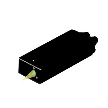
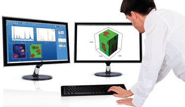
Raman Solutions Brochure - Find the Raman system best suited to your applications
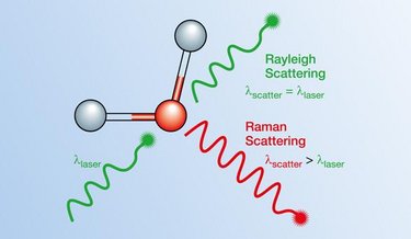
Our Raman instruments are designed to deliver unparalleled performance, offering detailed insights into sample characterization. Whether you're in academia, industry, or research, our Raman spectrometers provide the appropriate spectral resolution, real-time monitoring, and user-friendly software to meet your analytical needs.