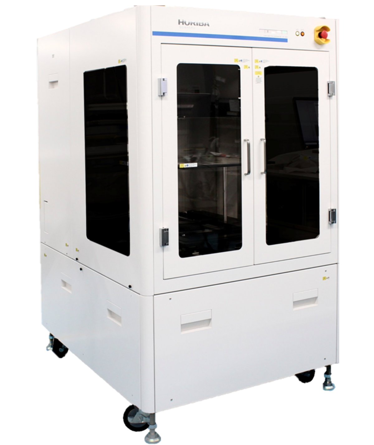
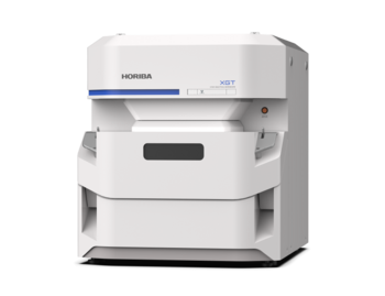
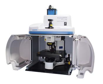
Designed to revolutionize large sample analysis and expand your applications
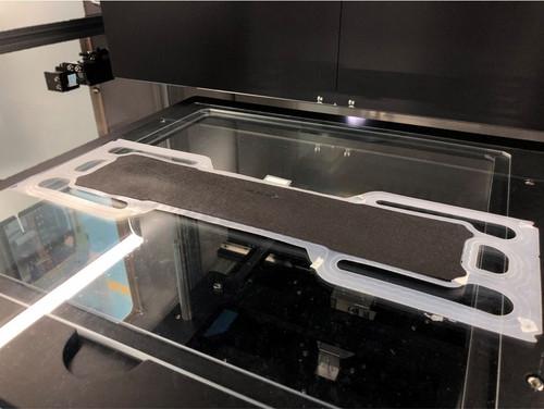
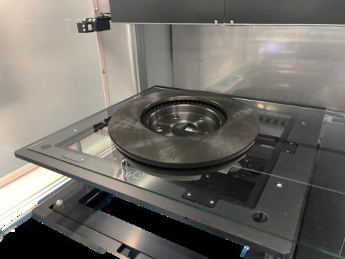
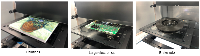
The XGT-9000SL series has a super large chamber capacity up to 1030 mm x 950 mm x 500 mm [W x D x H] and includes a wide stage by 500 mm x 500 mm. The features enable users to analyze even sizable samples such as paintings, large printed circuit boards, and brake rotors. It is perfect for handling such large samples with ease and carried out elemental analysis on it non-destructively. It is also versatile to accommodate multiple small sized samples on the stage to carry out sequential analysis.
Dual types of detectors for fluorescent X-rays and transmission X-rays
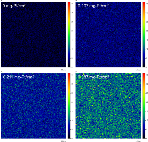
The XGT-9000SL Series provides dual types of detectors. One is a fluorescent X-ray detector which tells the elemental distribution of a sample, and the other is a transmission X-ray detector which tells the internal structure of a sample. XGT-9000SL can get the two types of images of the same area simultaneously. Especially it is helpful for a better understanding of electronics, gemstones including inclusions, and biological specimens.
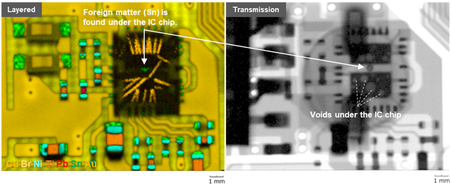
Simultaneous imaging of elemental image and transmission X-ray image of a printed circuit board
(Left) Elemental layered image revealed a foreign matter stuck under the IC chip. (Right) Transmission X-ray image revealed many voids under the IC chip.
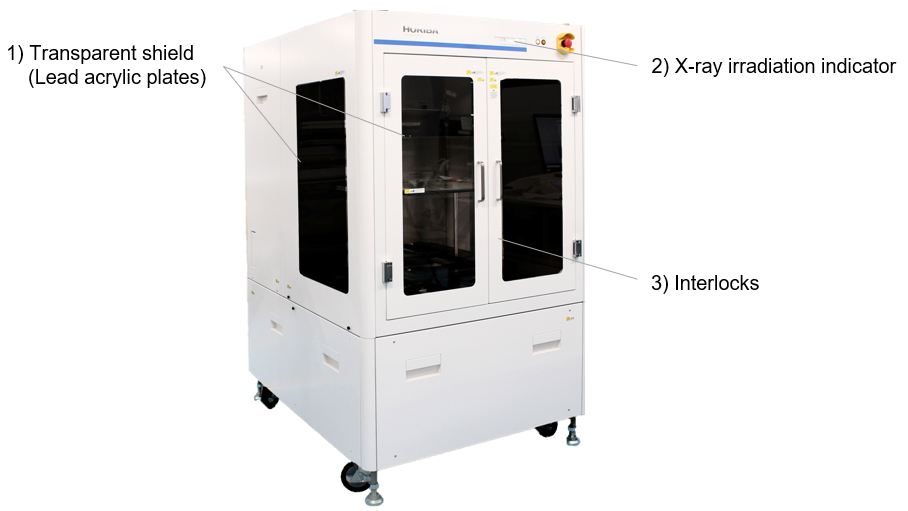
Protect users from X-ray exposure with the X-ray shield chamber
The XGT-9000SL is designed with highest level of X-ray shielding against X-ray radiations (IEC-61010-1 / JAIMAS0101-2001 compliant). With its 1) transparent shield, users can easily observe and monitor the sample in the chamber during a measurement without compromising user safety. The system also includes 2) an X-ray irradiation indicator located on the front panel alerting users when X-rays are active. Furthermore, the XGT-9000SL is equipped with 3) interlocks on the chambers doors, preventing any X-ray irradiation in the unlikely event that an user accidentally opens the door while the system is in operation.
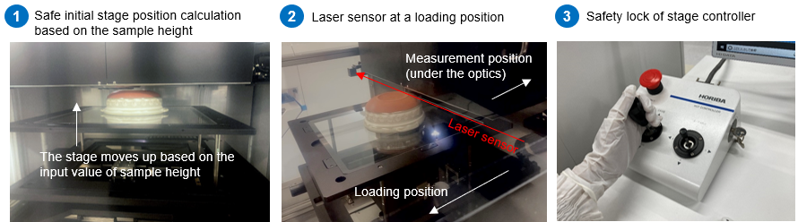
Multiple safety functions protect your precious samples from collision risks
Multiple safety functions are incorporated to ensure the protection of your precious samples:
- Stage position calculation: The software can calculate the safe initial position based on an input value of a sample height. This means that the stage will move up and stop at the optimal position, minimizing the risk of accidental collisions with the optics or other sensitive components.
- Laser sensor: The system can detect interference from your samples with a laser sensor and stop the stage before your sample contacts with the optics even if an inaccurate sample height has been input.
- Stage lock: The XGT-9000SL Series comes equipped with a sophisticated stage controller that incorporates a safety stage lock function. Even if an user accidentally operates the stage in a wrong way, the stage will not go beyond the designated safe position.
Flexible and user-friendly software
The XGT-9000SL Series software has a highly flexible and user-friendly interface. It displays data trees, optical images, spectrum results, the periodic table, and map imaging results at a glance. Users also have the flexibility to customize the on-screen layout, or even to show the results on multiple monitors. Thus, it allows users to see the results more clearly and boosts your data analysis.
Apart from the standard software functions, the advanced modules can be added to the software suite to provide more comprehensive user experiences.
- Multilayer FPM module: for thickness measurement with/without standard samples
- RoHS module: for RoHS screening
- Queue module: for automated multiple measurements in unattended mode
- Particle Finding module: for particle analysis and co localized analysis
- LabSpec Link module: for data transfer to LabSpec 6 for multivariate analysis
Application Examples