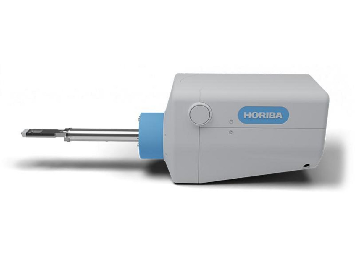
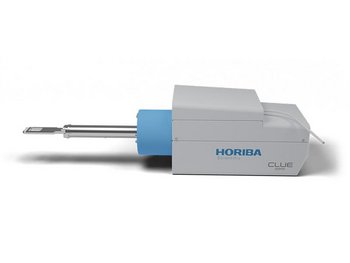
R-CLUE

Cathodoluminescence R-CLUE MM Picture Fr

Raman Photoluminescence & Cathodoluminescence

The R-CLUE all-in-one fiber coupled solution offers colocalized Raman, PhotoLuminescence and Cathodoluminescence Imaging and spectroscopy analysis.
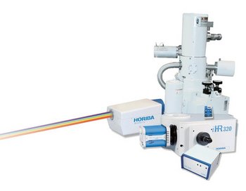
R-CLUE takes benefit of HORIBA Scientific extended know-how in Raman and PL spectroscopy with field-proven optical components, guaranteeing optical stability, reproducibility and collection efficiency.

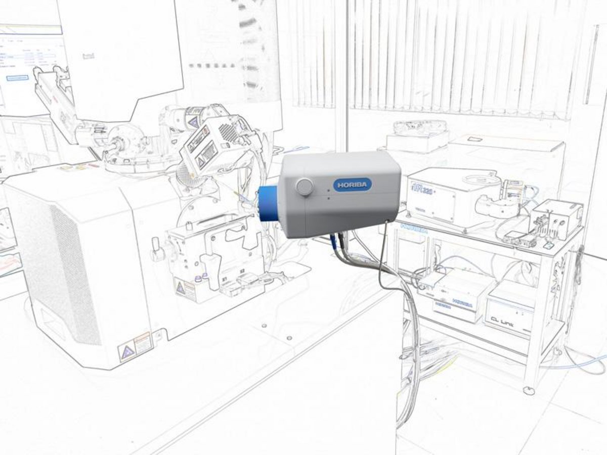
Thanks to its compact footprint inside and outside the SEM  and its deported spectroscopy modules, it can be installed on a large range of specimen chambers.

R-CLUE is fully automated and computer controlled, from retractable parabolic mirror position to Rayleigh filter change and grating and detector selection, allowing quick adoption by even non spectroscopist  users.

Controlled by Labspec 6 Simply Powerful software suite, R-CLUE features advanced unique functionalities for both fast imaging and spectroscopy analysis (KIA spectral database, SWIFT, cosmic ray removal, peak fitting, particle finder, mosaic, multivariate analysis, advanced 3D display…)

Segment: Scientific
Fabricant: HORIBA France SAS

All-in-one fully automated solution for high-end Research labs, featuring:

  • Fiber coupled interface
  • Colocalized laser and e-beam excitations
  • Fully computer controlled
  • Multi-wavelengths Raman lasers
  • Injection-rejection Raman filters
  • Field upgradeable
  • Built-in video camera

Spectral Range

UV-VIS or VIS-NIR fiber dependent

Diamond turned collection mirror

parabolic mirror
short & long working distances

200 mm retractable interface

Motorized with fine adjustment under vacuum

CL imaging detector

Standard: Panchromatic and monochromatic CL with ambient PMT
Options: Cooled PMT, IGA monochannel, photon counting PMT, time resolved PMT, RGB filters

CL spectrometer type

iHR 320, iHR 550 (320 to 550 mm focal length),up to 3 grating turret, 2-entrance/2exit

Upgrade available

Upgrade from LabRAM HR Series

Electron beam control

CL-LINK for multiple acquisition processing (Analog, pulse mode, SE), Mapping linescan,
point measurement, Synchronization with spectroscopic detection, Control by external scan input of Electron Microscope

Software

Spectroscopy and imaging powered by LabSpec 6™

Remote Controller

Optional

Clue Accessories

Polarization, ND filter, camera, EMCCD etc

SEM Accessories

LN2, He cryo-stages, EBIC detector and many other accessories to complement our CLUE Series add-on detectors

Defect Evaluation of GaN Epitaxial Wafer by CL
The threading dislocation occurs easily in GaN crystal grown on sapphire substrates. It is said that this is caused by the large lattice mismatch of sapphire and GaN. The crystal may seem to be uniform in the SEM image, but the dark spot such as the threading dislocation can be observed when measuring the CL intensity image at the wavelength (362nm) which corresponds to the band edge emission.
Luminescent Defects in Synthetic CVD Diamond Films Localized by Cathodoluminescence Spectroscopy
The characterization of synthetic CVD diamond material by hyperspectral cathodoluminescence spectroscopy and imaging allows the detection and accurate location of the promising NV luminescent point defects for innovative solid-state quantum mechanical systems. In this work we performed CVD epitaxial growth on a pattern of micro-pillars etched on a diamond substrate. Cathodoluminescence (CL) analysis revealed that NV centres were successfully localized at the edges of the pillars.
The Evaluation of Phosphor for White LEDs by CL Image
Phosphor plays a key role to obtain white light as for blue LED + phosphor and near ultraviolet LED + phosphors. In order to improve high bright white LEDs, it is necessary that the whole phosphor particle emits light homogeneously. When measuring with CL the phosphor used as white LED, the area which does not emit light in phosphor particle can be observed. CL system is used for evaluation of non-luminescent area to improve luminescent efficiency and characteristics.

Request for Information

Do you have any questions or requests? Use this form to contact our specialists.

* These fields are mandatory.

Related products

Cathodoluminescence - CLUE Series

Cathodoluminescence Solutions for Electron Microscopy

F-CLUE
F-CLUE

Compact Hyperspectral Cathodoluminescence

H-CLUE
H-CLUE

Versatile Hyperspectral Cathodoluminescence

Cathodoluminescence - CLUE Series

Cathodoluminescence Solutions for Electron Microscopy

F-CLUE
F-CLUE

Compact Hyperspectral Cathodoluminescence

H-CLUE
H-CLUE

Versatile Hyperspectral Cathodoluminescence