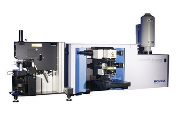


Figure: An Optical Microscope Observation System
Static Image Analysis uses high-resolution microscopy images of particles mounted on a slide or filter to measure particle size and shape. Because the particles remain stationary, the same particles can also be analyzed with complementary techniques for chemical or elemental identification.
Analyze the Same Particle with Multiple Methods
Static Image Analysis offers a unique advantage because the same particle remains stationary and can be analyzed by multiple methods. Because the particles remain stationary on a glass slide or filter, the technique goes far beyond mere optical particle size and shape analysis - it allows for targeted analysis using techniques such as lasers or X-rays to perform molecular structure and elemental analysis on the exact same particles.
Component-specific Particle Size Distributions
In mixed samples where multiple components coexist, identifying the chemical properties of individual particles allows you to output not just the particle size distribution of the entire sample, but independent particle size and shape distributions for each specific component.
Targeted and Efficient Analysis
Prior morphological information can be used to filter particles efficiently. For example, chemical analysis can be triggered only for particles within a specific size range or for particles with irregular shapes.
Accurate Identification of Foreign Matter and By-products
Even when particles look visually identical, chemical and elemental analysis can distinguish target components from foreign matter or by-products, enabling statistical evaluation of their sources and potential impact.
The following techniques may be used depending on the imaging source and the type of signal being measured.
It is also useful to understand the imaging platform, since the available size range and sample requirements depend strongly on how images are acquired. Particle Image Analysis uses different imaging methods depending on the particle size range and the information needed. Two common approaches are optical microscopy and electron microscopy. Electron microscopes provide much higher resolution than optical microscopes because electrons have a shorter wavelength than visible light. However, they generally require the sample to be placed in a vacuum. In contrast, optical microscopes typically have a lower resolution limit of around 0.5 µm, but they can be used on a wider range of sample states and are well suited for many Particle Image Analysis applications.
While Static Image Analysis provides highly detailed particle data, it faces two main practical challenges: sample preparation and locating the exact same particle across multiple instruments.
Static Image Analysis requires particles to be evenly distributed on a substrate without overlapping. Manual dispersion can cause agglomeration, which reduces measurement accuracy and reproducibility. To address this, automated dispersion systems using pressure differentials are commonly used to spread dry powders uniformly and minimize operator-induced errors.
HORIBA Solution: XD-100 Particle Disperser
When combining different analytical methods such as Raman and SEM/EDX, finding the exact same micron-sized particle on a second instrument can be technically difficult and time-consuming. This issue can be addressed using coordinate transfer systems or reference markers on the sample substrate. These tools transfer absolute XY coordinates and angular orientation between microscopes, allowing efficient relocation and analysis of the target particle.
HORIBA Solution: nanoGPS navYX
The Two Paradigms of Image Acquisition
In Static Image Analysis (SIA), data acquisition generally falls into two operational categories: wide-field imaging, which captures broad areas simultaneously, and point-by-point scanning, which builds surface information sequentially. While optical microscopy excels at rapidly capturing wide fields of view in a single exposure, advanced techniques such as Raman spectroscopy and X-ray analytical microscopy inherently rely on scanning mechanisms to acquire detailed chemical or elemental data.
The Challenge of Full-Area Mapping
When analyzing particulate samples, a significant challenge arises regarding time and resource efficiency. The analytical focus is strictly on the particles themselves, rather than the substrate or background plate upon which they are dispersed. Consequently, conducting a comprehensive, full-area mapping scan across the entire surface—including the empty spaces between particles—is highly inefficient and unnecessarily prolongs the total measurement time.
The Smart Software Solution: Targeted Analysis
To resolve this bottleneck, advanced software solutions have been developed to intelligently optimize the analytical workflow. In scanning analytical instruments equipped with an optical microscopy function for sample observation and positioning, a highly effective approach is utilized. The software first uses a rapid, wide-field optical microscope image to automatically detect and map the exact coordinates of the particles on the substrate. Once these regions of interest are identified, the system exclusively targets and analyzes only the specific areas where particles are present.
By ignoring the empty background, this software integration significantly reduces overall measurement time, improves instrument availability, and maximizes the relevance of the acquired data.
Static Image Analysis measures particles immobilized on a slide or filter, enabling precise focusing and very high-resolution shape evaluation. Because the particles remain stationary, it also supports correlative analysis on the exact same particles using probes such as lasers or X-rays, which is difficult for fast-moving particles in dynamic or in-line/on-line setups.
Static Image Analysis calculates particle size distributions and detailed shape parameters by extracting particle outlines from high-quality images. It can also combine optical morphology with chemical/molecular/elemental identification on the same particles through targeted irradiation (e.g., lasers or X-rays).
By identifying the chemical properties of individual particles, Static Image Analysis can report not only the overall size distribution, but also independent size and shape distributions for each component in a mixed sample. This is especially valuable when multiple components coexist and morphology alone is not sufficient to distinguish them.
Two common challenges are (1) sample preparation—particles must be evenly dispersed on a substrate without overlapping, and manual preparation may not sufficiently break up agglomerated material or achieve consistent dispersion, which can compromise measurement accuracy and reproducibility—and (2) relocating the exact same micron-sized particle across instruments for correlative workflows. These are typically addressed using automated dispersion approaches (e.g., pressure-differential dispersion) and coordinate transfer systems or reference markers that share XY coordinates and orientation between microscopes.
Yes. Because particles remain stationary on a slide or filter, Static Image Analysis enables analysis of the exact same particles and supports targeted follow-up measurements (e.g., Raman via laser irradiation, or elemental analysis), with relocation commonly supported by coordinate transfer systems or reference markers.
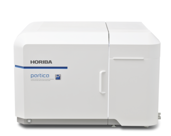
Laser Diffraction and Dynamic Imaging Particle Size and Shape Analyzer
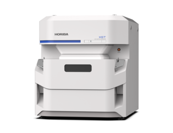
X-ray Analytical Microscope (Micro-XRF)
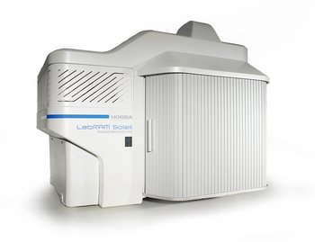
Raman Spectroscope - Automated Imaging Microscope
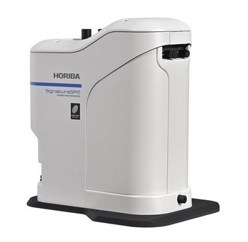
Scanning Probe Microscope with Chemical Signature
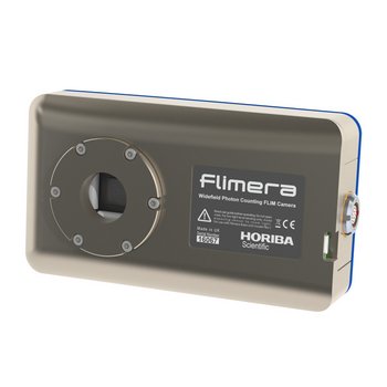
SPAD array imaging camera for dynamic FLIM studies at real time video rates
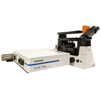
Fluorescence Lifetime Imaging Platform
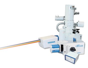
Cathodoluminescence Solutions for Electron Microscopy
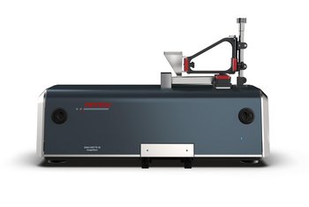
Dynamic Image Analysis
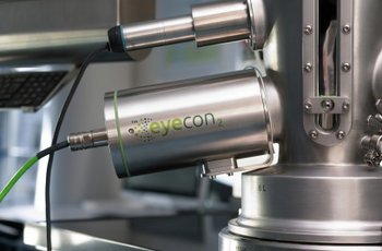
Direct Imaging Particle Analyzer
Tiene alguna pregunta o solicitud? Utilice este formulario para ponerse en contacto con nuestros especialistas.