Measuring Modes
Contact AFM in air;
Contact AFM in liquid (optional);
Semicontact AFM in air;
Semicontact AFM in liquid (optional);
True Non-contact AFM;
Dynamic Force Microscopy (DFM, FM-AFM);
Dissipation Force Microscopy;
Top Mode;
Phase Imaging;
Lateral Force Microscopy (LFM);
Force Modulation;
Conductive AFM (optional);
I-Top mode (optional);
Magnetic Force Microscopy (MFM);
Kelvin Probe (Surface Potential Microscopy);
Single-pass Kelvin Probe;
Capacitance Microscopy (SCM)
Electric Force Microscopy (EFM);
Single-pass MFM/EFM (“Plane scan”);
Force curve measurements;
Piezo Response Force Microscopy (PFM);
PFM-Top mode;
Nanolithography;
Nanomanipulation;
STM (optional);
Photocurrent Mapping (optional);
Volt-ampere characteristic measurements (optional).
Measuring Modes in liquid with AFM Head HE001 and Liquid cell
Contact AFM;
Semicontact AFM;
TopMode;
Phase Imaging;
Lateral Force Microscopy (LFM);
Force Modulation;
Force curve measurements;
Nanolithography;
Nanomanipulation.
Scanner and Base
Sample scanning range: 100 µm x 100 µm x 20 µm (±10 %)
Scanning type by sample: XY non-linearity 0.05 %; Z non-linearity 0.05 %
Noise: 0.1 nm RMS in XY dimension in 100 Hz bandwidth with capacitance sensors on; 0.02 nm RMS in XY dimension in 100 Hz bandwidth with capacitance sensors off; < 0.1 nm RMS Z capacitance sensor in 1000 Hz bandwidth
Resonance frequency: XY: 7 kHz (unloaded); Z: 15 kHz (unloaded)
Base
Manual sample positioning: range 25x25mm, positioning resolution 1um;
Motorized SPM measuring head positioning: 1.6x1.6mm, positioning resolution 1um;
Motorized approach: 1.3 mm;
Sample holder for standard slides and cover glasses;
Optional Sample holder: Maximum sample size: 50.8x50.8 mm, 5 mm height with capability to choose measuring area 25x25mm in any quadrant of 50.8x50.8 mm area or in center of sample.
AFM Head HE001
Laser wavelength: 1300nm;
No registration laser influence on biological sample;
No registration laser influence on photovoltaic measurements.
Registration system noise: <0.03nm.
Fully motorized: 4 stepper motors for cantilever and photodiode automated alignment.
Free access to the probe for additional external manipulators and probes;
Top and side simultaneous optical access: planapochromat objectives 10x, NA=0.28 and 20x, NA=0.42 respectively.
Liquid cell (optional)
Manual sample positioning: range 15x15mm, positioning resolution 1um;
Holder for Petri dish dia.35mm;
Volume of liquid: 1.5-2.5 ml;
Flow-through for liquid exchange: two tubes.
Conductive AFM unit (optional)
Current range: 100fA ÷ 10uA;
3 current ranges (1nA, 100na and 10uA) switchable from program;
Voltage range: -10 ÷ +7V;
RMS current noise: less than 60fA for 1nA range.
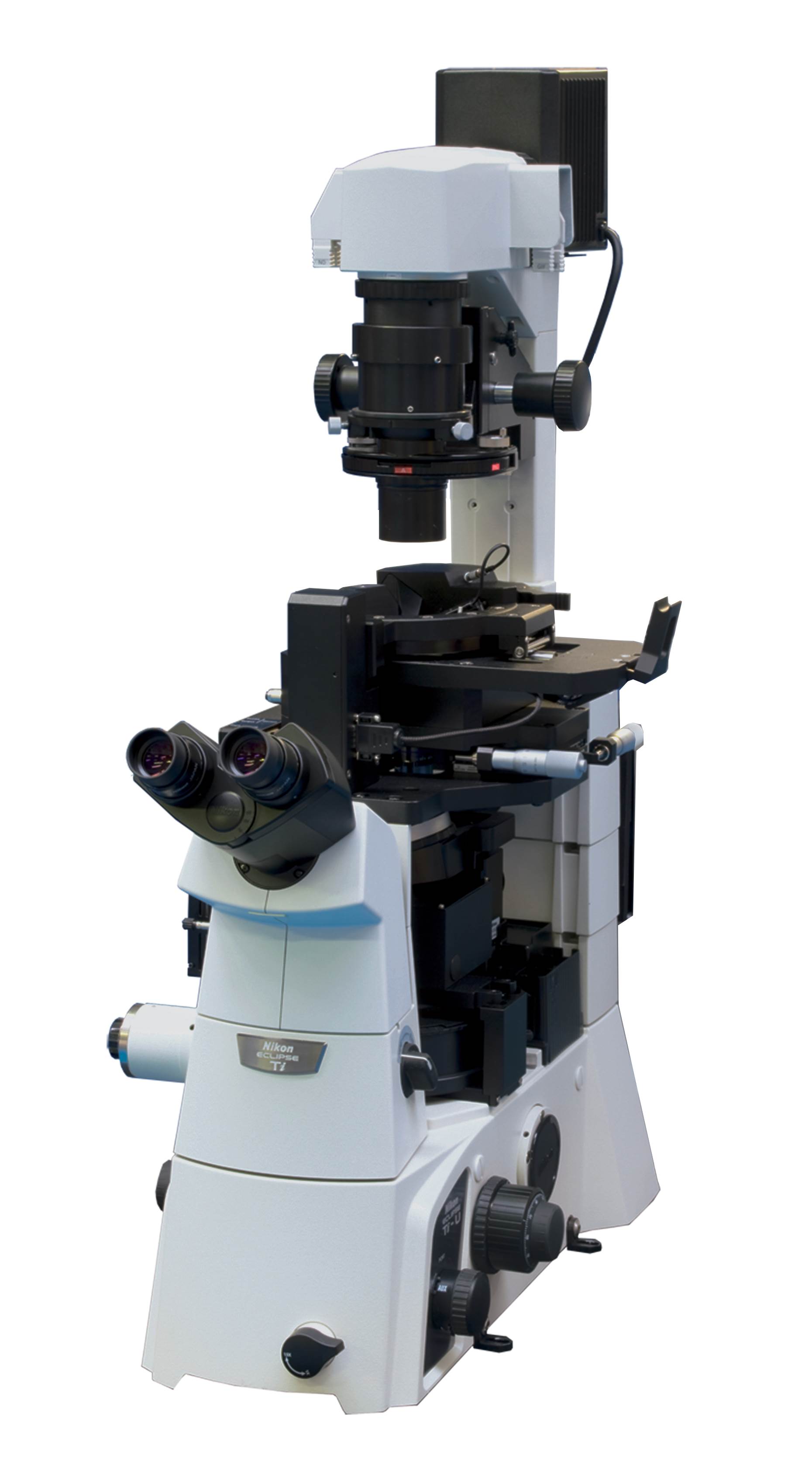
Compatibility with inverted optical microscopes
No interference with optical imaging due to infrared laser;
Capability to install on:
Nikon Ti-E, Ti-U, Ti-S, TE2000;
Olympus IX-71, IX-81;
Phase contrast, DIC and fluorescent techniques with native optical condenser;
Upgradeability to TRIOS platform for spectroscopic and TERS operation.
Optical microscope for standalone operation (optional)
Numerical aperture: up to 0.1;
Magnification on 19" monitor with 1/3" CCD: from 85x to 1050x.
Horizontal field of view: from 4.5 to 0.37 mm
Manual detent zoom: 12.5x
Stand and coarse/fine focusing unit.;
Capability to use planapochromat objectives: 10x, NA=0.28 and 20x, NA=0.42 and 100x, NA=0.7 (depends on AFM head);
Software
Automatic alignment of registration system;
Automatic configuration and presetting for standard measuring techniques;
Automatic cantilever resonance frequency adjustment;
Capability to work with force curves;
Macro language Lua for programming user functions, scripts and widgets;
Capability to program controller with DSP macro language in real time without reloading control software;
Capability to process images in coordinate space including making cross-sections, fitting and polynomial smoothing up to 8 degree;
FFT processing with capability to treat images in frequency space including filtration and analysis;
Nanolithography and nanomanipulation;
Processing up to 5000x5000 pixel images.


