Size:
2.96
MB
LabSpec 6 Brochure
Contrôle et affichage de volumes confocaux 3D
La cartographie Raman de volumes confocaux 3D d'échantillons offre une véritable vue chimique des structures internes grâce à l'interrogation chimique à haute résolution spatiale limitée par la diffraction que procure la microscopie Raman.
Accédez à notre centre de ressources vidéo LabSpec 6 pour maîtriser parfaitement votre logiciel de spectroscopie LabSpec 6.
En général, l'expérience permet d'acquérir un ensemble de plans 2D selon l'axe Z de l'échantillon, avec un spectre Raman complet associé à chaque pixel du volume 3D XYZ obtenu.
Le module avancé d'affichage de surfaces et de volumes 3D (3D Surface and Volume Display) de LabSpec 6 permet une interrogation complète de ces données de volume grâce à des fonctions de rotation 3D, de filtrage, de transparence et de tranchage. L'éclairage réglable permet d'ajuster la luminosité d'éléments spécifiques du volume. Un mode « immersif » en option permet de visualiser l'image sous tous les angles et selon toutes les orientations possibles, même depuis l'intérieur du volume.
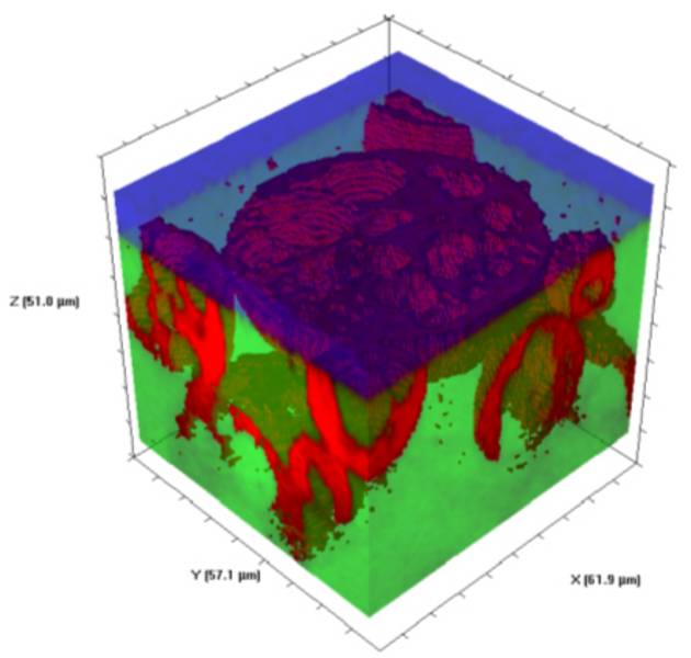
Volume 3D d'une bille de polymère expansé dans une matrice de polymère. L'image montre la bille, la matrice et l'huile d'immersion [données fournies par Neil Everall, Intertek Wilton, R.-U.].
Le module d'affichage de surfaces et de volumes 3D permet également d'obtenir un rendu 3D complet d'images de surface 2D, ce qui convient parfaitement à l'affichage d'images de topographie AFM avec superposition d'informations chimiques Raman. L'utilisateur exerce un contrôle total sur la rotation de l'image, la mise à l'échelle et l'éclairage réglable. Une option de mode « immersif » permet de visualiser l'image selon toutes les orientations et sous tous les angles possibles.
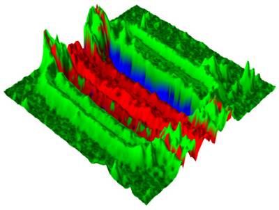
Vue de surface 3D complète d'une structure en polysilicium, illustrant l'intensité totale du silicium (surface) avec la répartition du polysilicium, du silicium cristallin et du silicium nanocristallin.
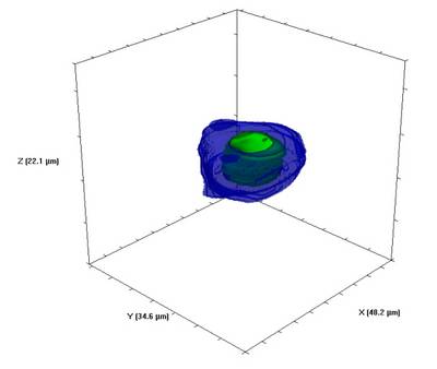
Vue du volume 3D d'une inclusion géologique, montrant l'emplacement du dioxyde de carbone (CO2) en phase gazeuse et aqueuse dans une matrice de quartz. La matrice n'est pas représentée.
Do you have any questions or requests? Use this form to contact our specialists.