
PDF
0.58
MB

HORIBA新型微区XRF XGT-9000对玻璃填充红宝石的表征
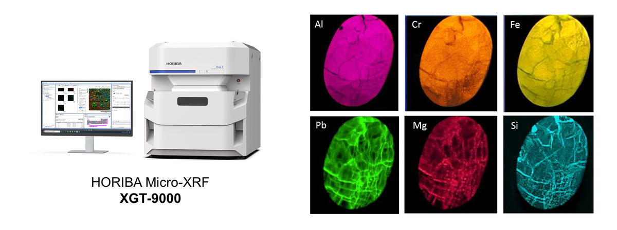
用于首饰的宝石通过加热、辐射或填充裂纹等工艺来处理,可使其更加完美。我们使用XGT-9000 微区X射线荧光分析仪对Mineralab公司提供的红宝石进行了成像分析。通过透射X射线成像结果,可发现红宝石内部存在部分缺陷,而荧光X射线成像则显示出红宝石表面经过填充处理,其中包括Pb、Mg和Si等代表铅玻璃的物质。

Lead-glass-filled rubies are treated gemstones in which lead-glass is introduced into fractures that reach the surface to improve their appearance. To understand the extent and distribution of lead-glass filling, it is important not only to confirm the presence of Pb (lead), but also to visualize its distribution within the gemstone.
X-ray fluorescence spectrometer (XRF) can non-destructively confirm the presence of Pb, but it doesn’t provide elemental distribution information. LA-ICP-MS offers highly sensitive elemental analysis; however, it involves minor surface damage caused by laser ablation. Elemental mapping is possible but requires scanning the sample while progressively removing material from the surface.
In contrast, micro-XRF elemental imaging generates spatial distribution maps of Pb without damaging the gemstone, allowing the extent of lead-glass filling along fractures to be visualized intuitively.
Figure 1. Comparison of XRF, LA-ICP-MS, and Micro-XRF
Micro-XRF can visualize not only Pb but also multiple elements within the gemstone. By comparing elemental imaging, additional information about the gemstone and its treatment can be obtained. For detailed analytical results, please refer to the application note below.
Figure 2. HORIBA micro-XRF XGT-9000 system (left) and elemental distribution images of a glass-filled ruby (right)
如您有任何疑问,请在此留下详细需求信息,我们将竭诚为您服务。