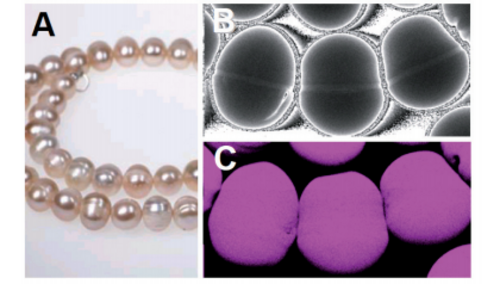
Wear metals and additive Elements Analysis.

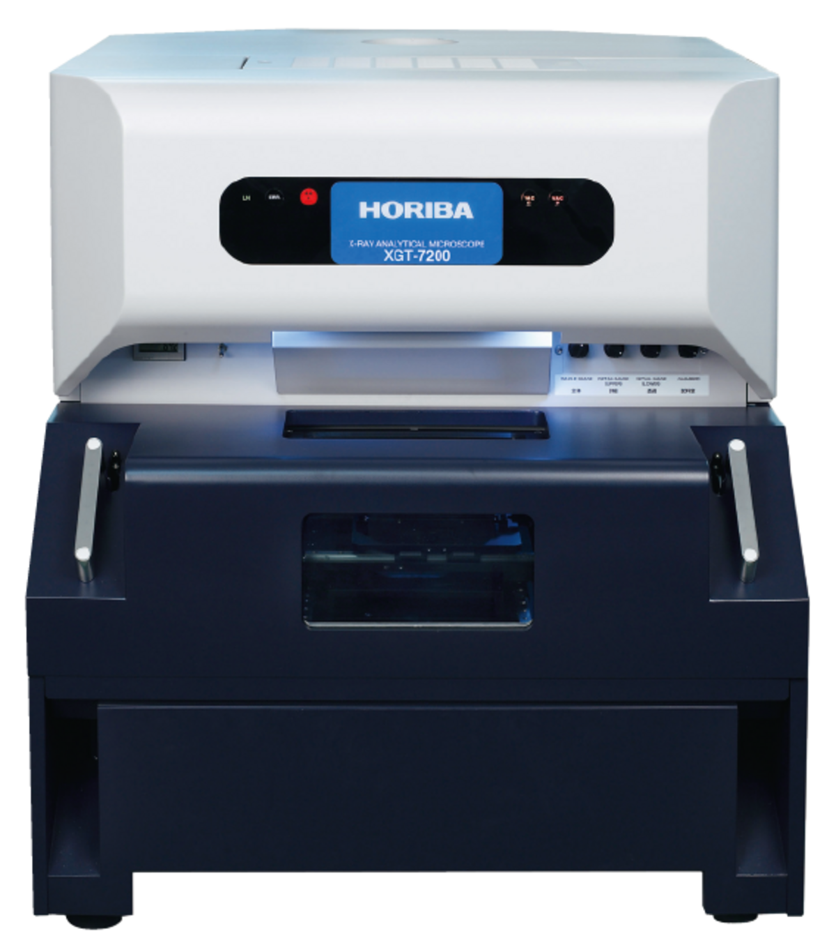
 This product has been discontinued and is no longer available. You can still access this page for informational service purposes.
真空チャンバ装備により軽元素の高感度分析が可能
用途:異物 分析、故障 解析、不具合 解析、RoHS 対応、スクリーニング、非破壊 分析、食の安全、鉛フリー、規制、鑑識、生態分析、生体分析
対象:プリント基板、実装基板、電子部品、家電、玩具、製薬、医薬品、食品、めっき
機能:マッピング、面分析、微小部 分析
■デュアル バキューム チャンバ
元素マップを極めるスマートマップ
サンプルから得られる元素情報を全て保存。
一度スキャンしてしまえばサンプルがなくてもマップ像の追加や点・面分析も可能に。また、マッピングスピードも高速のため短時間分析を実現。
ナビゲータウィンドウにより操作手順をサポート
分析手順を知らなくてもナビゲータウィンドウによって分析手順がわかり、そのフロー(ボタン)を選択するだけで目的の操作ができます。各部分での操作は充実したアドバイスにより簡単にわかり、精度の良い自動定性と定性確認機能で、正確な分析が行なえます。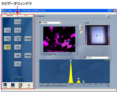
軽元素の感度向上
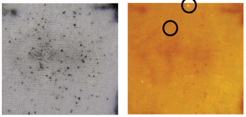
●隕石の分析例
Si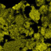 | Mg |
軽元素(Mg,Siなど)でも真空チャンバを使うことにより高感度な測定を実現。もちろん従来通り、大気中軽元素測定を可能とする真空プローブ機構も搭載しています。
 | 薬剤 ・錠剤内部、薬剤に混入した異物の分析等 ・取り出し、断面出し等前処理なしで分析可能 |  | 自動車 ・PM(粒子状物質) ・塗料、エンジンオイル中の異物等 ・多点分析で複数位置も一度で測定 |
 | 地質 ・岩石、堆積物の分析 ・風化状態の確認等 ・分断しにくい大きな試料も そのままセッティング可能 |  | 材料 ・合金、金属、半導体、コンクリート等 ・成形済みの各種成品の元素分析 |
 | バイオ ・植物や動物などの生体試料 ・歯、臓器などの生体組織試料 ・含水試料も分析可能 |  | エレクトロニクス ・樹脂モールド中の不良解析 ・ICパッケージの不良解析 ・コンデンサ、LED等の不良解析 ・非破壊で故障解析が可能 |
 | 法科学 ・塗膜片、インク成分の判定 ・金属無機材、有機材の判定 ・異同識別の手法 |  | 考古学 ・陶磁器の染付け ・ガラス玉(トンボ玉)、勾玉 ・絵画の顔料 ・貴重な考古学試料の元素分析 |
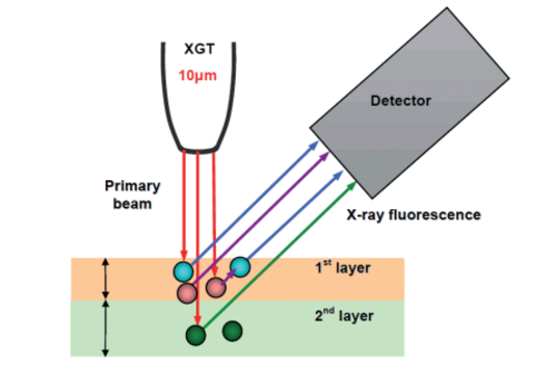
測定原理 | エネルギー分散型蛍光X線分析 |
|---|---|
測定元素 | Na(ナトリウム)〜U(ウラン) |
分解能 | 10 µm/100 µm |
最大測定エリア | 100×100 mm |
設置寸法(本体) | 670 (W)×820 (D)×760 (H) mm |